
Patella Fractures
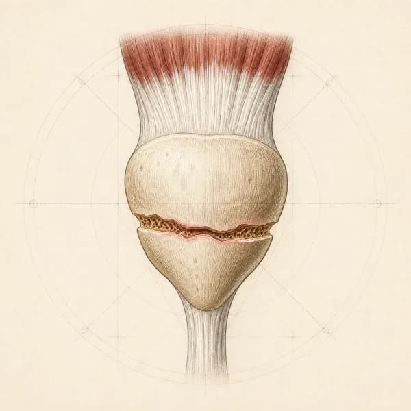
A patella fracture is a break in the kneecap, often after a fall or direct blow. It can cause pain, swelling and difficulty straightening the knee. A suspected fracture needs medical assessment first. Rehabilitation follows the treating team's instructions for weight-bearing, knee movement and protection, whether or not surgery is needed.
Key points
- What usually helps
- Following the protection plan from your doctor or surgeon, then rehab once healing allows.
- What you can do now
- Follow your team's protection plan. Keep the thigh muscle switching on.
- When to see a doctor first
- Some signs need emergency care. Check the list before you start treatment.
Symptoms
Does this sound like you?
- Pain and swelling at the front of the knee after a fall or a direct blow
- Difficulty straightening the knee or lifting the straight leg
- Tenderness over the kneecap itself
- Bruising over the front of the knee
- Pain when trying to put weight on the leg
Usually follows a fall onto the knee or a blow to the front of the knee. The key question is whether you can still straighten the knee against gravity. That tells your doctor whether the extensor mechanism (the thigh muscle, its tendons and the kneecap, which together straighten the knee) is intact.Book an assessment for patella fractures
When to see a doctor first
Go to emergency
An open wound over the kneecap after the injury
Go to emergency now: an open fracture needs urgent treatment to prevent infection
Calf pain, swelling or warmth, or sudden shortness of breath or chest pain, especially while in a brace or walking less
Possible blood clot. Calf symptoms need same-day medical assessment; shortness of breath or chest pain means go to emergency now
Severe pain, tightness or numbness in the leg that keeps getting worse, especially in a cast or brace
Go to emergency now to rule out pressure building in the leg (compartment syndrome)
Get seen today
You cannot straighten the knee or lift the straight leg after a fall or blow to the kneecap
Possible fracture that has broken the system that straightens the knee (the extensor mechanism). Go to emergency or an urgent care clinic the same day for an X-ray
After surgery: a wound that is red, hot, leaking or increasingly painful, or a fever
Possible infection. Contact your surgical team the same day, or go to emergency
A new click, clunk or sudden loss of the ability to straighten the knee during rehab
Possible failure of the fixation or the healing bone. Stop the exercises and contact your surgeon the same day
Treatment
How it is treated
A typical rehabilitation progression
Three phases, from settling symptoms to returning to full activity.
Recovery from patella fractures is usually staged: calm the symptoms first, then rebuild the strength and capacity of the area, then return to your full activities. The phases below show how that can progress.
Phase 1Protect the Fracture and Wake Up the Quadriceps (Weeks 0 to 6)
The early phase depends on whether the fracture was treated with or without surgery, and on the surgeon's protocol. Either way, the bone needs to be protected, and the front thigh muscle (quadriceps) needs every chance not to shut down completely. I work inside the brace settings and weight-bearing instructions you have been given, and I do not push knee bending, or straightening the knee against resistance, beyond what the surgical team has cleared.
Example exercises
- Tightening the thigh muscle without moving the knee ( quad sets) in the brace with the knee straight, 10 to 15 repetitions with 5-second holds, several times a day
- Straight leg raises in the brace, if cleared by the surgeon, 3 sets of 10, focusing on keeping the knee locked fully straight
- Ankle pumps (pointing the foot up and down) and calf work to reduce swelling and keep blood moving in the lower leg
- Gentle knee bending, moved for you or with help, within the surgeon's allowed range, often starting around 30 to 60 degrees and increasing week by week
- Pain and swelling control with ice, elevation, and compression as appropriate
Possible progression milestones
The thigh muscle clearly switching on, with a visible contraction and a without the knee sagging (no lag), pain and swelling going down, and clearance from the surgical team to increase knee movement and weight-bearing in phase 2.
Phase 2Rebuild Range, Strength, and Confident Weight-Bearing (Weeks 6 to 16)
Once the bone is stable on examination and the surgeon has cleared progression, the focus shifts to getting the knee's movement back, rebuilding the front thigh and the muscles around the hip, and returning to putting your full weight through the leg. This phase often covers weaning off the brace. I watch for pain that comes on later after sessions, because it is one of the more sensitive signs that the load is building too fast.
Example exercises
- Thigh exercises with the foot on the ground (closed-chain), built up gradually, starting with shallow squats against a wall and mini-squats, 3 sets of 10 to 12
- Step-ups and step-downs onto a low step, progressing height as control allows, 3 sets of 8 to 10 per side
- Stationary bike once the knee bends enough to pedal a full turn comfortably, starting at very low resistance and increasing the range over time
- Strengthening the muscles on the side and back of the hip ( and extensors) with band work and bridges, 3 sets of 10 to 15, to share load through the whole leg
- Single-leg balance on firm ground, moving to foam as your sense of joint position () improves, 3 sets of 30 to 45 seconds
Possible progression milestones
Full or nearly full pain-free knee movement, a controlled single-leg squat part of the way down without the knee dropping inward, thigh strength clearly improving on repeated testing, and going up and down stairs comfortably, one foot per step.
Phase 3Return to Running, Sport, and Impact Work (Months 4+)
The final phase turns a stable, strong leg into one that can handle impact, slowing down and the demands of the activities you care about. Progress depends on meeting criteria, not on dates. I use measurable benchmarks for strength, and for hop results matching between the legs, before clearing contact or high-speed sport, and I accept that some people will take six months and some closer to a year.
Example exercises
- Heavier strengthening with the foot on the ground, such as split squats, Bulgarian split squats (back foot on a bench) and moderate-load squats, 3 sets of 6 to 10
- Straight-line running, building from walk-jog intervals to steady running and then faster tempo runs, watching for any pain at the front of the knee the day after
- Agility and change-of-direction work starting with low-intensity cutting drills and progressing toward sport-specific patterns
- A set of hop tests (single hop, triple hop and crossover hop) to compare sides, used as a checkpoint for moving on rather than just a measure of results
- Sport-specific reintroduction layered in only once strength and hop symmetry benchmarks are met, rather than on a fixed calendar
Possible progression milestones
Front thigh strength close to the uninjured side on strength testing (isometric or isokinetic), matching results between the legs on the hop tests, confidence with sport-specific slowing down and cutting, and no meaningful pain or joint swelling the day after sessions as the load builds.
What you can do now
- Follow your team's protection plan. Your weight-bearing, how far the knee may bend and your brace settings are set by your treating team, whether or not you had surgery. Follow those over any general timeline, including the one on this page.Do not start straight-leg raises or loaded exercises until your surgeon has cleared them and you can lift the leg without the knee sagging.
- Keep the thigh muscle switching on. Once your surgeon is satisfied the fracture is stable, tightening the thigh muscle without moving the knee is usually safe and helpful. It helps keep the thigh muscle from shutting down and the knee from stiffening while the bone heals.
- Keep swelling down and blood moving. Ankle pumps (pointing the foot up and down) help reduce swelling and keep blood moving in the lower leg. Ice, raising the leg and compression can also help control pain and swelling, as appropriate.
- Stop if you feel a new click or clunk. A new click or clunk, or suddenly losing the ability to straighten the knee during rehab, can mean the fixation or the healing bone has failed. Stop the exercises and contact your surgeon the same day.
- Know the other warning signs. Calf pain, swelling or warmth needs a same-day medical assessment, and sudden shortness of breath or chest pain means go to emergency now. Severe pain, tightness or numbness in the leg that keeps getting worse, especially in a cast or brace, also means go to emergency now.After surgery, a red, hot, leaking or increasingly painful wound, or a fever, means contact your surgical team the same day, or go to emergency.
What I use in clinic
Similar conditions
Conditions that can feel similar
- Quadriceps Tendon Rupture
- A gap above the kneecap rather than at the bone, and being unable to straighten the knee yourself, can suggest a quadriceps tendon rupture. The kneecap may sit lower than usual. It needs urgent assessment, because kneecap height alone is not a diagnostic test. It often happens when the thigh muscle is suddenly loaded as it lengthens, in an older person or someone with risk factors that weaken tendons.
- Patellar Tendon Rupture
- A gap that can be felt below the kneecap, being unable to straighten the knee, and a kneecap that sits high on an X-ray. Typical in younger, athletic people after a sudden jump or landing, and on examination the extensor mechanism no longer straightens the knee.
- Patellar Dislocation or Subluxation
- A twisting injury, or one that pushes the knee inward (valgus), with the knee giving way. The person becomes apprehensive when the kneecap is pushed outward on examination, and the tissue along the inner edge of the kneecap (the medial retinaculum) is tender. The kneecap itself is usually intact on imaging, though an injury to the cartilage and underlying bone (osteochondral injury) of the inner kneecap or the outer end of the thigh bone can occur.
- Bipartite Patella
- A variation in how the kneecap develops, in which one part, usually the upper outer corner, forms as a separate piece of bone (a separate ossification centre). It is often found by chance on imaging. There is typically no sudden injury, the edges are rounded and dense (sclerotic) rather than sharp, and the extensor mechanism is intact.
- Osteochondral Injury of the Patella or Trochlea
- Sudden pain and swelling inside the joint after a twisting or direct injury, with catching or locking, but no clear fracture line on a standard X-ray. MRI shows the cartilage surface and any loose fragment in the joint (loose body).
- Prepatellar Bursitis
- A clearly outlined, soft, fluid-filled swelling in front of the kneecap, rather than tenderness over the bone or a problem with the extensor mechanism. There is often a history of repeated kneeling rather than a sudden hard impact, and you can still straighten the knee yourself.
Questions
What patients ask me most
How does a patella usually get fractured?
Two main ways. The first is a direct blow to the front of the knee, for example a fall directly onto the kneecap. The second is an indirect overload, where the thigh muscle (quadriceps) contracts hard while the knee is bending and pulls the kneecap apart. Direct blows tend to break the kneecap into several pieces (a comminuted fracture), while indirect overload tends to cause a break straight across (a transverse fracture). Both can disrupt the extensor mechanism, the muscle, tendons and kneecap that straighten the knee, which is the main thing that decides whether surgery is needed.
How is the decision made between a cast or brace and surgery?
The short answer is whether the extensor mechanism still works and whether the bone pieces have moved out of place (displaced). Melvin and Mehta's 2011 review in the Journal of the American Academy of Orthopaedic Surgeons describes the common criteria for treatment without surgery: pieces that have not moved, or have barely moved, an intact extensor mechanism (the patient can lift the straight leg or straighten the knee against gravity), and a step in the joint surface (articular step-off) of roughly 2 to 3 mm or less. Fractures that do not meet those criteria generally go to surgery to fix the bone, because the knee joint surface and the extensor mechanism matter for long-term function.
What does non-operative treatment look like day to day?
Typically a period in a brace locked with the knee straight or nearly straight, protected weight-bearing as tolerated depending on the surgeon's guidance, and gradually bringing back knee bending and thigh muscle work over weeks rather than days. Gwinner and colleagues' 2016 GMS Interdisciplinary Plastic and Reconstructive Surgery DGPW overview is useful here: the priority is protecting the healing bone while not letting the quadriceps completely shut down or the knee stiffen permanently. Early tightening of the thigh muscle without moving the knee (isometric quadriceps contractions) and straight leg raises are usually safe and helpful once the surgeon is satisfied the fracture is stable.
Why is the quadriceps the focus of rehab after a patella fracture?
Because the kneecap is a bone that sits inside the quadriceps tendon (a sesamoid bone), and the whole job of the extensor mechanism is to straighten the knee against gravity and control it as it bends during walking, going down stairs and sport. After a fracture, the thigh muscle often switches off (inhibition) and starts to lose bulk (atrophy) within days. Getting it working again, then strong, then able to absorb and produce force through the knee, is what rebuilds a confident, working leg. Without that work, people plateau with a stiff and weak knee even if the bone itself has healed.
Can I expect to get back to sport?
Many people do, but the timeline is long and the criteria matter more than the calendar. Typical return-to-sport criteria after a patella fracture include full, pain-free knee movement, front thigh (quadriceps) strength within about 10 to 15 percent of the uninjured side on measured testing, similar results on both legs in single-leg tests such as a single-leg squat and a series of hop tests, and confidence in sport-specific movements. Rushing back before those benchmarks risks re-injury or lasting pain at the front of the knee. I build the rehab around the criteria, not the weeks.
Will I have arthritis in this knee later?
The risk is higher than for an uninjured knee, particularly when the joint surface was disrupted or a step remains in it. Gwinner and colleagues note that osteoarthritis behind the kneecap after injury (post-traumatic patellofemoral osteoarthritis) is a recognised long-term outcome after patella fracture, more so when the pieces could not be lined up perfectly or the injury was severe. That is one reason good early management matters. It is also why ongoing quadriceps strength and sensible loading through the years ahead is worth the time, because it directly protects the joint.
When can I start bending the knee again?
That is a surgeon-led decision and depends on the fracture pattern, whether it was fixed surgically, and how stable the repair is. In broad terms, light, protected bending is often introduced within the first few weeks after surgical fixation, or once a fracture treated without surgery is stable on examination. Bending hard and early on a fragile repair can make the fixation or the bone fail. I follow the operating surgeon's specific protocol rather than a generic template, and I communicate with that team when timing is unclear.
Do I still need physiotherapy if the fracture was undisplaced?
Often, yes. Even without surgery, a patella fracture can switch off the quadriceps and stiffen the knee during the protected phase. Rehabilitation can help restore movement, strength and confidence once the fracture is ready for each step. The amount of support needed depends on the injury, the treatment and how you progress.
Guides on knee pain
Longer explanations of symptoms and scan-report wording.
- Pain Right Below the KneecapA single tender spot below the kneecap that flares with jumping, stairs, or deep squats. Most commonly patellar tendinopathy in active adults, and growth-plate conditions in young athletes.
- Suprapatellar Effusion (Fluid on the Knee)Extra fluid inside the knee joint, seen in the pouch above the kneecap. What the size words on a report mean, the usual causes by how quickly the swelling came on, and when to see a doctor first.
- Joint Space Narrowing in the KneeThe gap between the bones looks thinner than expected on an X-ray. What medial, tricompartmental and the severity words mean, how it relates to pain, and what the guidelines recommend.
Related Conditions
Conditions I commonly see alongside, or confused with, this one.
Patellofemoral Pain (Kneecap Pain)
Pain around the kneecap can continue after the fracture has healed
Patellar Tendinopathy (Jumper's Knee)
Both affect the front of the knee and the extensor mechanism
Knee Osteoarthritis
A fracture into the joint surface raises the risk of osteoarthritis later
PCL Injuries
Both commonly follow a direct blow to the front of the knee, such as a dashboard injury
ACL Injuries
Both need a staged return to running and cutting once the knee has healed
MCL/LCL Sprains
Getting back to it
Physiotherapy for patella fractures, built around the activity you want back.