Knee Pain
e.g., Patellofemoral Pain Syndrome, Patellar Tendinopathy
Overview
The Science of Knee Pain
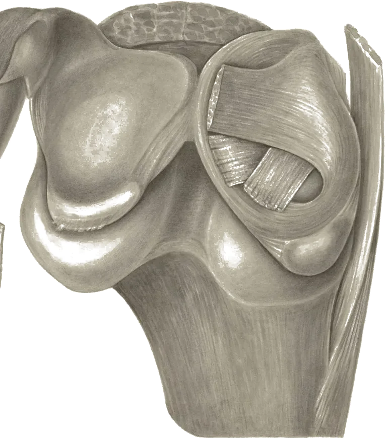
Link copiedpain syndrome involves dysfunction at the interface where your kneecap (patella) meets your thigh bone (femur). The condition typically develops when your patella doesn't track properly in its groove during knee movement, creating abnormal pressure and stress on the joint cartilage.
The patella normally glides smoothly in the trochlear groove of the femur, but when muscle imbalances or structural factors alter this tracking, certain areas of cartilage experience increased compression. This can lead to irritation of pain-sensitive structures including the joint capsule, synovium, and subchondral bone.
Research shows that people with patellofemoral pain often have altered during functional activities, with changes in how forces are distributed across the joint during weight-bearing movements like squatting, stair climbing, and landing from jumps.
Overview
Contributing Factors
Link copiedYour patella acts like a pulley to increase the mechanical advantage of your quadriceps muscles. When this system isn't working optimally, several biomechanical factors contribute to the problem.
Hip weakness, particularly in the and gluteus maximus, often allows your thigh to collapse inward during activities. This creates a angle at the knee that pulls your patella laterally, increasing stress on the lateral facet.
Quadriceps imbalances, especially weakness in the vastus medialis oblique (VMO) relative to the vastus lateralis, can contribute to poor patellar tracking. The VMO is crucial for pulling the patella medially and preventing lateral drift.
Your foot mechanics also play a role - excessive can create internal rotation of the tibia, which alters the angle of pull from your quadriceps and affects patellar tracking. Tight structures like the , lateral , or hip flexors can create additional forces that pull the patella out of optimal alignment.
Symptoms
Clinical Presentation
Link copiedPrimary Symptoms
Associated Symptoms
Typical pattern
I typically see pain that develops gradually without a specific injury. It's often worse with activities that load the knee in flexion - my patients frequently tell me about pain climbing stairs, getting up from sitting, or after long car rides. The pain tends to be more noticeable during and after activity rather than at rest.
Symptoms
Differential Diagnosis
Link copiedConditions with similar presentations:
Patellar Tendinopathy (Jumper's Knee)
Key differences: Pain localized to the inferior pole of the patella rather than around or behind the kneecap. Reproduced by single-leg decline squat and resisted knee extension. Typically aggravated by jumping, landing, and sprinting rather than by prolonged sitting or stairs.
Meniscal Pathology
Key differences: Joint-line tenderness, often with a history of a twisting injury or deep squatting event. True mechanical symptoms like catching, locking, or giving way are more common. Positive McMurray or Thessaly tests support the diagnosis, and symptoms are not dominated by prolonged sitting.
Early Tibiofemoral Osteoarthritis
Key differences: Typically older patient group with morning stiffness lasting under 30 minutes, , and pain along the medial or lateral joint line rather than around the patella. Stiffness after periods of rest improves with movement, and imaging may show joint space narrowing.
Iliotibial Band Syndrome
Key differences: Sharp lateral knee pain near the lateral femoral epicondyle, not around the patella. Commonly presents in runners and cyclists, often at a consistent distance or cadence. Tenderness over the and pain with repetitive knee flexion around 30 degrees help differentiate.
Medial Plica Syndrome
Key differences: Medial knee pain with a palpable, tender band over the medial femoral condyle. Often gives a snapping or clicking sensation with knee flexion and extension. Symptoms may mimic pain but are more focal and reproducible with direct palpation of the plica.
Referred Pain from the Hip
Key differences: Anterior knee pain with limited or painful hip internal rotation and a or FABER test that reproduces knee symptoms. Knee examination is often unremarkable. More common in hip or femoroacetabular , and should be screened in any anterior knee pain presentation.
When to seek professional help
Research
Key Research & Evidence
Peer-reviewed studies supporting the treatment approach.
Finding
Combined hip and knee exercise produces superior pain reduction compared to knee exercise alone
Research details
A 2022 systematic review and meta-analysis of 65 RCTs published in Journal of Orthopaedic & Sports Physical Therapy found hip-and-knee-targeted exercise therapy showed standardized mean difference (SMD) of 1.02 (95% CI: 0.58, 1.46) for pain and SMD of 1.03 (95% CI: 0.61, 1.45) for function at 3 months compared to knee-targeted exercise alone, while knee-targeted exercise showed SMD of 1.16 (95% CI: 0.66, 1.66) for pain versus control
Clinical relevance
In the 2022 JOSPT review (Neal et al., 65 RCTs), hip-and-knee-targeted exercise therapy showed efficacy for pain and function compared with knee-targeted exercise alone at 3 months, with moderate to large effect sizes. This supports including hip strengthening as a core component of patellofemoral pain rehabilitation rather than treating it as an optional adjunct
Finding
Hip strengthening produces greater pain reduction effect than knee strengthening
Research details
A 2025 systematic review and meta-analysis by Zhang and colleagues in the European Journal of Medical Research (30:90) comparing hip versus knee strengthening training in patellofemoral pain found hip-focused protocols were associated with a larger pain reduction effect (standardized mean difference, SMD -1.740) than knee strengthening (SMD -1.302), where more negative values indicate greater pain reduction
Clinical relevance
Hip strengthening targeting external rotators and abductor muscles produces superior and faster pain relief compared to quadriceps-only approaches, supporting hip-dominant or equal hip-knee exercise prescription in early rehabilitation phases to optimize symptom resolution and functional recovery
Finding
6 interventions show positive effects at 3 months but long-term evidence lacking
Research details
The 2022 JOSPT meta-analysis by Neal and colleagues (65 RCTs) identified 6 interventions with positive effects at 3-month follow-up: knee-targeted exercise therapy, combined interventions, foot orthoses, lower-quadrant manual therapy, hip-and-knee-targeted exercise therapy, and knee-targeted exercise therapy combined with perineural dextrose injection, though no intervention was adequately tested beyond 3 months, leaving long-term efficacy uncertain
Clinical relevance
While multiple interventions demonstrate short-term efficacy at 3 months, lack of long-term data beyond this timepoint limits ability to guide extended rehabilitation planning, suggesting need for ongoing exercise maintenance programs beyond initial 3-month intervention period to prevent symptom recurrence
Management
Evidence-Based Management
Treatment strategies with the strongest support in the current literature.
Primary approach
Hip and knee strengthening exercises targeting , external rotators, and quadriceps achieve significant pain reduction and functional improvement in pain
Complementary
Movement retraining with feedback and patellar taping provide additional benefits when combined with exercise therapy to address biomechanical factors
Prevention & long-term
Proper training progression and neuromuscular control exercises may help reduce the risk of pain in active individuals and athletes
Detailed management strategies
Activity Modification
Temporarily reducing aggravating activities allows tissues to settle while maintaining fitness through alternative activities
Important precautions
- Avoid complete rest
- Monitor for symptoms spreading to other areas
Progressive Loading Program
Gradual increase in activity helps build tissue tolerance and addresses underlying weakness patterns
Important precautions
- Progress based on symptoms
- Ensure proper form before increasing intensity
Movement Awareness
Learning to recognize and modify movement patterns that aggravate symptoms helps prevent flare-ups
Important precautions
- Focus on quality over quantity
- Seek guidance if unsure about techniques
Pain Monitoring
Understanding your pain patterns helps guide activity levels and identifies when to seek additional help
Important precautions
- Persistent increase in pain warrants reassessment
- Night pain is not typical
Management
Treatment Techniques
Evidence-based manual therapy and intervention approaches.
Treatment approaches supported by current research and clinical guidelines
Recommended treatment approaches
Treatment approaches are individualized to each patient's needs and goals. All interventions require explicit informed consent, and treatment plans are collaboratively modified based on your preferences and response to care.
Rehabilitation
A Typical Rehabilitation Progression
Three phases, from settling symptoms to returning to full activity.
Recovery from Knee Pain is usually staged: calm the symptoms first, then rebuild the strength and capacity of the area, then return to your full activities. The three phases below show the kind of progression the evidence supports and that I commonly work through in clinic. They are here to show you what the road can look like, not to act as a personal program.
- Phase 1
Symptom Settling and Foundational Strength
Reduce irritability, restore tolerance to daily loading like stairs and sitting, and begin isolated hip and quadriceps strengthening. This follows the 2019 JOSPT Clinical Practice Guideline, which gives strong evidence for combined hip and knee exercise as first-line care.
Examples, not a prescription
- Side-lying , 3 sets of 12 to 15, focusing on glute medius activation
- Short-arc quad extensions through a pain-free inner range, 3 sets of 10 to 12
- Wall sit at a shallow angle (under 45 degrees of knee flexion), 3 sets holding 20 to 45 seconds
- with slight hip external rotation, 3 sets of 10 to 12 per side
Ready to progress when
Pain under 3 out of 10 with daily activities like stairs and getting out of a chair, no increase in symptoms 24 hours after exercise, and the ability to perform prescribed exercises with clean technique.
- Phase 2
Progressive Loading and Movement Quality
Load the knee through deeper ranges with good mechanics, add closed-chain and single-leg work, and start addressing the collapse pattern that is often present. Movement retraining is informed by the JOSPT Clinical Practice Guideline and is integrated throughout this phase.
Examples, not a prescription
- Goblet squat to a box, 3 to 4 sets of 8 to 10, progressing depth and load as symptoms allow
- Step-ups and step-downs with cueing to prevent knee collapse, 3 sets of 8 to 10 per side
- Split squats or reverse lunges, 3 sets of 8 to 10 per side
- Single-leg Romanian deadlift for posterior chain and pelvic control, 3 sets of 8 per side
- Spanish squat or heavy slow resistance leg press for tendon and quadriceps loading, 3 sets of 8 to 10
Ready to progress when
Pain-free or minimal pain (under 2 out of 10) through full-depth squatting and single-leg work, good frontal-plane control on step-downs (no visible knee valgus), and tolerance for 2 to 3 strength sessions per week without flare.
- Phase 3
Return to Sport, Running, and Higher Loads
Reintroduce the specific demands of the patient's activity, whether that is running, cutting sports, or recreational training. Emphasis is on capacity, running mechanics, and a sustainable maintenance plan, since recurrence is common without ongoing loading (per the JOSPT CPG).
Examples, not a prescription
- Graded return-to-run program using walk-run intervals, progressing volume then intensity
- Running cadence work cued at a 5 to 10% increase to reduce load
- Bilateral then single-leg hop progressions for landing mechanics, 3 to 4 sets of 6 to 8
- Heavier bilateral squatting and deadlifting matched to the patient's training goals
- Sport-specific drills (cutting, change-of-direction, jumping) layered in once tolerance is confirmed
Ready to progress when
Full return to desired sport or activity with pain 2 out of 10 or less and no 24-hour flare, single-leg hop tests within 10 to 15% of the unaffected side, and a maintenance strengthening plan the patient can sustain at least twice weekly.
Management
Prognosis & Recovery
What outcomes and recovery factors typically look like.
Expected timeline
Most people see meaningful improvement in 6-12 weeks with appropriate exercise therapy. Full recovery typically takes 3-4 months
Natural history
Without appropriate treatment, symptoms often persist and can become chronic, potentially limiting participation in physical activities and affecting quality of life
Factors affecting recovery
Management
Measuring Progress
How to track the recovery arc week to week.
Day-to-day tracking
I track changes in your pain during specific activities like stairs and squatting, your ability to perform daily tasks without limitation, and improvements in movement quality during functional tests
Assessment tools
Anterior Knee Pain Scale to quantify functional limitations and monitor improvement over time
Activity targets
Return to your desired activities - whether that's running, sports participation, or simply navigating stairs without discomfort
Management
Frequently Asked Questions
Common concerns and answers about this condition.
Is walking good for patellofemoral pain?
Is walking good for patellofemoral pain?
Walking on flat ground is usually well tolerated and is one of the first activities I reintroduce. It keeps the knee moving without the high compressive loads of deep squats or downhill running. joint force is lowest near full extension and rises with deeper knee flexion, which is why stairs and prolonged sitting tend to flare symptoms more than walking does.
How long does patellofemoral pain take to heal?
How long does patellofemoral pain take to heal?
Most people improve meaningfully within 6 to 12 weeks of consistent exercise therapy, with full resolution often taking 3 to 4 months. The 2019 JOSPT Clinical Practice Guideline (Willy et al.) supports hip and knee strengthening as the most effective approach. Long-term follow-up studies, though, show a substantial proportion of people still have some symptoms years later if rehabilitation stops early or is inconsistent. That is why I treat maintenance strengthening as part of the plan, not an optional extra.
Do I need an MRI for patellofemoral pain?
Do I need an MRI for patellofemoral pain?
Usually not. pain is a clinical diagnosis based on history and examination, and imaging does not reliably correlate with symptoms. I consider MRI when symptoms fail to improve with appropriate rehab, when there is true locking or giving way, significant trauma, or signs pointing toward or ligamentous injury, not as a first-line investigation.
Why does my knee hurt going down stairs more than up?
Why does my knee hurt going down stairs more than up?
Descending stairs is an , single-leg task. Pooled data on healthy adults puts peak joint reaction force during stair descent at roughly 2.8 times body weight, higher than walking. If the quadriceps and hip musculature cannot control knee flexion well, that load concentrates on a smaller patch of cartilage behind the kneecap, which is a common driver of pain.
Can I keep running with patellofemoral pain?
Can I keep running with patellofemoral pain?
Often yes, with modifications. Reducing mileage, avoiding steep downhill running, and cueing a slightly quicker cadence (around 5 to 10% higher) can help. Heiderscheit and colleagues showed a 10% increase in step rate meaningfully reduces energy absorption at the knee. Bramah and colleagues (2019) reported that a 10 percent increase in step rate was associated with improved running kinematics and clinical outcomes in runners with pain at 3-month follow-up. The pain rule I use is that symptoms stay at or below 3/10 during the run and settle within 24 hours. Persistent flares mean the load resets.
Is patellofemoral pain the same as runner's knee?
Is patellofemoral pain the same as runner's knee?
The terms are used interchangeably, but runner's knee is a general label that can include pain, syndrome, and patellar . Patellofemoral pain specifically refers to pain around or behind the kneecap, usually aggravated by loaded knee flexion activities like squatting, stairs, or prolonged sitting. Sorting out which one is present changes the rehab plan significantly.
Will my knee get worse if I keep exercising?
Will my knee get worse if I keep exercising?
Ongoing exercise does not appear to cause joint damage in pain, and inactivity is linked with worse outcomes. The goal is modifying load, not avoiding it. A short-term increase in symptoms after training is acceptable if it settles within 24 hours. If symptoms are worsening week over week, intensity or volume gets reduced until the trajectory reverses.
Are knee braces or taping helpful for patellofemoral pain?
Are knee braces or taping helpful for patellofemoral pain?
They can be a useful short-term adjunct. The 2019 JOSPT guideline supports patellar taping combined with exercise for short-term pain relief. Taping or a soft sleeve helps some patients tolerate their rehab exercises. It is not a long-term fix. Strength and movement quality drive the sustained change.
Related Conditions
Conditions I commonly see alongside, or confused with, this one.
- Anatomically related
Patellar Tendinopathy (Jumper's Knee)
Both affect the patellofemoral region; biomechanical factors often overlap
- Biomechanically linked
IT Band Syndrome
Both common in runners; IT band tightness can affect patellofemoral tracking
- Biomechanically linked
Lateral Hip Pain & Gluteal Tendinopathy
Gluteal weakness contributes to both lateral hip pain and poor knee tracking
Commonly confused with
Side-by-side comparisons for patterns that often get mistaken for knee pain.