Low Back Pain
acute and chronic, mechanical, disc-related
Overview
The Science of Low Back Pain
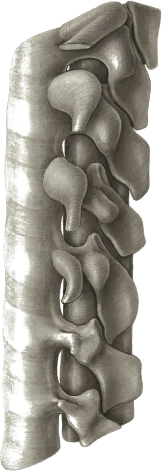
Link copiedMechanical low back pain typically involves dysfunction of the intervertebral discs, , , or surrounding musculature. The disc starts to lose its hydration and load distribution capabilities, which leads to increased stress on surrounding structures.
The deep stabilizing muscles like and transverse abdominis often show delayed activation patterns, compromising segmental stability. Over time, this can lead to movement pattern changes that perpetuate the problem.
When pain persists beyond 3 months, changes in the nervous system can amplify pain perception, making previously non-painful movements uncomfortable. When disc dysfunction progresses significantly, it may lead to disc with potential compression (sciatica). Similarly, when facet joints become primary pain generators, this can develop into facet joint syndrome, while sacroiliac joint dysfunction may become a distinct condition requiring specific treatment approaches.
Overview
Contributing Factors
Link copiedPoor posture and prolonged sitting create significant loads on your spine, particularly when you slouch or crane your head forward. Your core muscles - the deep abdominals and back extensors that act like an internal corset - often become weakened from inactivity, reducing the support they provide to your spine.
Heavy lifting with poor technique multiplies the forces through your discs. When you bend at the waist rather than squatting, you can increase bending stress on your discs, more so when bending is combined with twisting. Repetitive bending and twisting under load consistently exceed what the tissue can handle.
Modern lifestyle factors play a huge role: prolonged sitting, particularly with poor posture, increases disc pressure by approximately 30% compared to standing (though the difference is minimal with proper upright posture), and forward head posture from screen time changes how forces distribute through your entire spine. Even factors like tight hip flexors from sitting can alter your curve, forcing your back muscles to work overtime to maintain upright posture.
Symptoms
Clinical Presentation
Link copiedPrimary Symptoms
Associated Symptoms
Typical pattern
Pain typically worsens with sustained positions and improves with position changes. Morning stiffness improves with movement. Many people find certain movements consistently help or worsen their symptoms.
Symptoms
Differential Diagnosis
Link copiedConditions with similar presentations:
Lumbar Radiculopathy / Disc Herniation
Key differences: Leg pain dominant over back pain, often extending below the knee in a pattern. Positive or slump test, and sometimes specific weakness or reflex change. Worse with sitting, forward flexion, coughing, or sneezing.
Lumbar Spinal Stenosis
Key differences: Bilateral leg heaviness or cramping that builds with standing and walking and eases quickly with sitting or forward leaning (). Typically age over 60, with walking tolerance limited more by leg symptoms than by back pain itself.
Sacroiliac Joint Dysfunction
Key differences: One-sided pain localized just below the belt line at the PSIS, often pointed to with one finger. Reproduced by a cluster of provocation tests (distraction, thigh thrust, compression, Gaenslen, sacral thrust). Referral pattern rarely extends below the knee.
Facet-Mediated Pain
Key differences: Localized pain worse with extension and rotation, eased by forward flexion. Somatic referral into the buttock or posterior thigh typically stops at the knee. Neurological exam is unremarkable.
Hip Osteoarthritis (referring into low back)
Key differences: Groin or anterior thigh pain, reduced and painful hip internal rotation on examination, and worsening with weight-bearing rather than with positions. Low back symptoms are often secondary to altered gait and hip mechanics.
Vertebral Compression Fracture
Key differences: Acute focal spinal pain, often after low-energy event in someone older or on long-term corticosteroids. Pain is worse with loading and better lying down, and point tenderness over a single spinous process is common. Imaging is indicated when suspected.
Inflammatory / Serious Pathology (red flags)
Key differences: Unrelenting night pain, systemic features, unexplained weight loss, fever, history of cancer, or progressive neurological loss. These shift assessment from mechanical low back pain to an urgent medical review rather than physiotherapy alone.
When to seek professional help
Research
Key Research & Evidence
Peer-reviewed studies supporting the treatment approach.
Hayden JA, Ellis J, Ogilvie R, et al. · 2021
Exercise therapy for chronic low back pain
Cochrane Database of Systematic Reviews · n=24,486 participants (249 studies)
Key findings
Moderate-certainty evidence that exercise is probably effective for treatment of chronic low back pain compared to no treatment, usual care or placebo. Core strengthening, mixed exercises, and Pilates showed significant improvements.
Clinical relevance
Establishes exercise therapy as evidence-based first-line treatment with large body of supporting research
Hayden JA, Ellis J, Ogilvie R, et al. Exercise therapy for chronic low back pain. Cochrane Database Syst Rev. 2021;9(9):CD009790.
Wang XQ, Wang YL, Witchalls J, et al. · 2024
Physical therapy for acute and sub-acute low back pain: Expert consensus
Clinical Rehabilitation · n=22 international expert panel
Key findings
Strong evidence supports manual therapy combined with exercise for acute LBP. Multidisciplinary treatment more effective than single interventions for pain intensity reduction.
Clinical relevance
Provides current expert consensus on optimal physiotherapy approaches for acute presentations
Wang XQ, Wang YL, Witchalls J, et al. Physical therapy for acute and sub-acute low back pain: A systematic review and expert consensus. Clin Rehabil. 2024;38(6):715-731.
Kent P, Haines T, O'Sullivan P, et al. · 2023
Cognitive functional therapy for chronic, disabling low back pain (RESTORE trial)
The Lancet · n=492 participants
Key findings
Cognitive functional therapy produced large, sustained reductions in activity limitation at 13 weeks (mean difference 4.6 points on the RMDQ) and maintained the benefit at 52 weeks, compared with usual care. A movement-sensor biofeedback arm added no extra benefit.
Clinical relevance
Strongest modern evidence that an individualised biopsychosocial approach outperforms usual care for chronic disabling low back pain.
Kent P, Haines T, O'Sullivan P, et al. Cognitive functional therapy with or without movement sensor biofeedback versus usual care for chronic, disabling low back pain (RESTORE): a randomised, controlled, three-arm, parallel group, phase 3, clinical trial. Lancet. 2023;401(10391):1866-1877.
Fritz JM, Cleland JA, Childs JD · 2007
Treatment-based classification for low back pain
Journal of Orthopaedic & Sports Physical Therapy · n=Clinical commentary
Key findings
Outlines a treatment-based classification system that subgroups patients with low back pain by clinical presentation to match them to specific physiotherapy approaches, rather than applying a single generic protocol.
Clinical relevance
Supports individualized treatment approach based on clinical presentation rather than generic protocols
Fritz JM, Cleland JA, Childs JD. Subgrouping patients with low back pain: evolution of a classification approach to physical therapy. J Orthop Sports Phys Ther. 2007;37(6):290-302.
Research
Research Insights
Clinical implications and practice recommendations.
Exercise Therapy
Moderate-certainty evidence from a Cochrane review of 249 studies shows exercise is probably effective for chronic low back pain compared with no treatment, usual care, or placebo, though the evidence was too heterogeneous to recommend a single optimal exercise type or dose (Hayden et al., 2021)
Treatment Sequencing
Studies demonstrate that starting with to reduce irritability, then progressing to exercise therapy, produces superior outcomes compared to exercise alone in acute presentations
Biopsychosocial Approach
Cognitive functional therapy addressing pain beliefs and movement fears can produce greater improvement in disability than a traditional biomedical approach (Kent et al., RESTORE trial, Lancet 2023)
Classification Benefits
Patients matched to treatment based on their clinical presentation (Treatment-Based Classification) can experience better outcomes than those given a generic exercise programme
Long-term Effectiveness
Exercise therapy benefits are maintained at 12 months, while injection-based treatments show no long-term advantage over placebo
Management
Evidence-Based Management
Treatment strategies with the strongest support in the current literature.
Primary approach
Specific core and trunk stabilization exercises consistently outperform passive care for reducing pain and improving function, and they lower the chance of another episode
Complementary
techniques provide immediate pain relief and improved mobility when combined with active exercise programs
Prevention & long-term
Movement education and workplace ergonomic modifications reduce the risk of future episodes by addressing underlying biomechanical factors
Detailed management strategies
Graded Activity Pacing
Starting at a comfortable level and gradually increasing prevents flare-ups while building tolerance
Important precautions
- Avoid complete rest beyond 2 days
- Monitor for increasing leg symptoms
Position Modification
Small changes in how you sit, stand, and sleep can significantly reduce strain on sensitive structures
Important precautions
- Changes should reduce not increase symptoms
Regular Movement
Frequent position changes and gentle movement prevent stiffness and maintain mobility
Important precautions
- Stay within comfortable range
- Quality over quantity
Management
Treatment Techniques
Evidence-based manual therapy and intervention approaches.
Treatment approaches supported by current research and clinical guidelines
Recommended treatment approaches
Treatment approaches are individualized to each patient's needs and goals. All interventions require explicit informed consent, and treatment plans are collaboratively modified based on your preferences and response to care.
Pain Education & Self-Management
Understanding pain science to reduce fear and improve movement confidence alongside active rehabilitation.
Dry Needling
Precise needle therapy targeting trigger points for effective pain relief and improved muscle function.
Exercise Therapy
Personalized exercise programs designed to restore strength, flexibility, and function.
Joint Mobilization
Graded techniques to restore joint movement and reduce stiffness.
Soft Tissue & Myofascial Therapy
Targeted hands-on techniques to address muscle tension, pain, and movement restrictions.
Trigger Point Therapy
Focused pressure techniques to address painful trigger points and reduce muscle pain.
Cupping Therapy
Technique using controlled suction to address muscle tension and localized pain.
Postural Assessment & Movement Strategies
Analysis of posture and movement patterns to develop adaptable positioning strategies.
Rehabilitation
A Typical Rehabilitation Progression
Three phases, from settling symptoms to returning to full activity.
Recovery from Low Back Pain is usually staged: calm the symptoms first, then rebuild the strength and capacity of the area, then return to your full activities. The three phases below show the kind of progression the evidence supports and that I commonly work through in clinic. They are here to show you what the road can look like, not to act as a personal program.
- Phase 1
Foundation: Calm the Tissue, Restore Comfortable Movement
The goal of the first few weeks is to reduce symptom spikes, rebuild confidence moving, and find loaded positions you tolerate. Early active management, education, and gentle graded exercise are consistently recommended by NICE NG59, the ACP 2017 guideline, and the JOSPT 2021 low back pain CPG (George et al.).
Examples, not a prescription
- Short, frequent walks (5 to 10 minutes) rather than one long walk, built up over the first 1 to 2 weeks
- Cat-camel and pelvis tilts on hands and knees, 10 slow repetitions, for segmental mobility
- Supine hook-lying diaphragmatic breathing with gentle abdominal bracing, 5 to 10 breaths, several times daily
- Dead bug and modified bird dog to re-introduce spine-neutral control, 2 sets of 6 to 8 per side
- Breaks from sustained sitting every 30 to 45 minutes with a short stand or walk
Ready to progress when
Resting pain 3/10 or less, ability to sit and stand for 30 minutes without a meaningful flare, and confidence performing basic daily tasks (dressing, driving short distances, getting in and out of bed) without guarding.
- Phase 2
Progressive Loading: Build Capacity
Once symptoms settle, the spine needs load to get more durable. This phase rebuilds hip, trunk, and posterior chain strength, reintroduces hinging and squatting, and progresses work to the level of the patient's real-world demands. Exercise therapy is the most consistently supported intervention in the Hayden et al. 2021 Cochrane review.
Examples, not a prescription
- Hip hinge progressions: broomstick hinge, kettlebell deadlift, single-leg Romanian deadlift, 3 sets of 6 to 10
- Goblet squat to a box, progressed in depth and load, 3 sets of 8 to 10
- Glute bridge and single-leg bridge variations, 2 to 3 sets of 8 to 12
- Side plank and modified Pallof press for lateral and anti-rotation trunk control
- Farmer carries 20 to 40 metres for trunk stiffness under load, 3 to 4 rounds
Ready to progress when
Tolerating a moderate hinge and squat load with pain under 3/10, walking 30 minutes comfortably, and a full workday without symptom escalation.
- Phase 3
Return to Function: Demand-Specific Loading and Resilience
The last phase is about matching capacity to the job, sport, or hobby the person is going back to. For a construction worker that looks different than it does for a runner or a lifter. Building a maintenance plan the patient can keep doing is what protects against the high recurrence rate in the first 12 months described in the Lancet Low Back Pain Series.
Examples, not a prescription
- Trap bar or conventional deadlift, progressively loaded, 3 to 4 sets of 3 to 5
- Front squats or split squats at working loads, matched to patient goals
- Loaded carries and suitcase carries at moderate to heavy loads
- Return to running or sport built through a graded walk-run or low-impact framework
- A two- or three-session weekly maintenance programme the patient can sustain independently
Ready to progress when
Full work, home, and recreational demands with minimal pain, independent management of minor flares, and a written maintenance plan with realistic load targets.
Management
Prognosis & Recovery
What outcomes and recovery factors typically look like.
Expected timeline
Most acute episodes improve significantly within 2-6 weeks. Chronic patterns take 3-6 months for meaningful change
Natural history
Without addressing underlying factors, recurrence is common but doesn't mean worsening damage
Factors affecting recovery
Management
Measuring Progress
How to track the recovery arc week to week.
Day-to-day tracking
I track what changes day to day: pain interference with key tasks, movement quality during functional tests, and your confidence with daily activities
Assessment tools
Condition-specific questionnaires when useful (like the Oswestry for back pain or DASH for shoulder conditions)
Activity targets
One activity target that matches your goal - whether that's returning to sport, work tasks, or daily activities without limitation
Management
Frequently Asked Questions
Common concerns and answers about this condition.
Do I need an MRI for low back pain?
Do I need an MRI for low back pain?
Usually not in the first 6 weeks. NICE NG59 and the American College of Physicians guidance both advise against early imaging for non-specific low back pain because scans frequently show disc bulges, changes, and protrusions in people with no pain at all. Imaging becomes useful when there are red flags, progressive neurological loss, or when symptoms have not responded to a reasonable trial of conservative care and a scan would actually change the plan.
Should I rest or stay active?
Should I rest or stay active?
Stay active within tolerance. Bed rest beyond a day or two slows recovery and deconditions you.
How long does low back pain usually take to settle?
How long does low back pain usually take to settle?
Most acute episodes improve meaningfully in 2 to 6 weeks. The Lancet Low Back Pain Series describes a pattern of rapid early improvement followed by a slower tail, with many people experiencing some residual or recurring symptoms. Recurrence does not mean your back is getting worse. It usually reflects load, sleep, stress, and training history more than structural change.
What position should I sleep in?
What position should I sleep in?
The position that lets you sleep. There is no single correct posture. Side-lying with a pillow between the knees and back-lying with a pillow under the knees are the two setups that work for most people in the acute phase. If a position wakes you up sore, try the other.
Is it safe to keep exercising or lifting weights?
Is it safe to keep exercising or lifting weights?
Often yes, with short-term modifications. Completely stopping training tends to make things worse, not better. What I usually adjust first is deadlift volume, heavy loaded flexion, and deep morning bending, while keeping most of your programme intact. Pain during exercise up to about 3 or 4 out of 10 that settles within 24 hours is generally safe, and is closer to what the JOSPT 2021 low back pain CPG describes as appropriate loaded progression.
Why does my back feel worse in the morning?
Why does my back feel worse in the morning?
Discs rehydrate overnight, which makes the slightly taller and stiffer first thing, and mechanically more sensitive to bending in the first hour. Avoiding deep forward flexion (tying shoes bent over, loaded rowing, sit-ups) for 30 to 60 minutes after waking often takes the edge off. Prolonged, dramatic morning stiffness that lasts more than an hour and improves through the day, especially in a younger person, is a pattern I flag for inflammatory back pain screening.
My MRI showed disc degeneration. Is my back damaged?
My MRI showed disc degeneration. Is my back damaged?
Probably not in the way that report sounds. Brinjikji and colleagues (AJNR 2015) pooled imaging findings in pain-free adults and showed disc in around 37% of 20 year olds and 96% of 80 year olds, with disc bulges present in roughly 30% and 84% respectively. Those changes are part of a normal aging spine, not a structural diagnosis. I treat the clinical picture, not the scan.
When should I be worried about back pain?
When should I be worried about back pain?
Seek urgent care for loss of bladder or bowel control, numbness in the saddle area, rapidly progressing leg weakness, or foot drop. These can signal . Also see your physician promptly for unexplained weight loss, fever with back pain, severe unrelenting night pain, or a history of cancer, rather than starting physiotherapy first.
Related Conditions
Conditions I commonly see alongside, or confused with, this one.
- Anatomically related
Sciatica
Both involve the lumbar spine; disc dysfunction can progress to nerve root compression
- Common co-occurrence
Disc Herniations / Bulges
Disc problems often underlying cause of mechanical low back pain
- Biomechanically linked
Sacroiliac (SI) Joint Dysfunction
SI joint dysfunction commonly coexists with lumbar spine issues due to shared load transfer