MCL/LCL Sprains
Medial and lateral collateral ligament injuries
Overview
The Science of MCL/LCL Sprains
Link copiedMCL and LCL sprains involve stretching or tearing of the knee's . MCL injuries are more common, often from stress. These ligaments provide side-to-side stability.
Overview
Contributing Factors
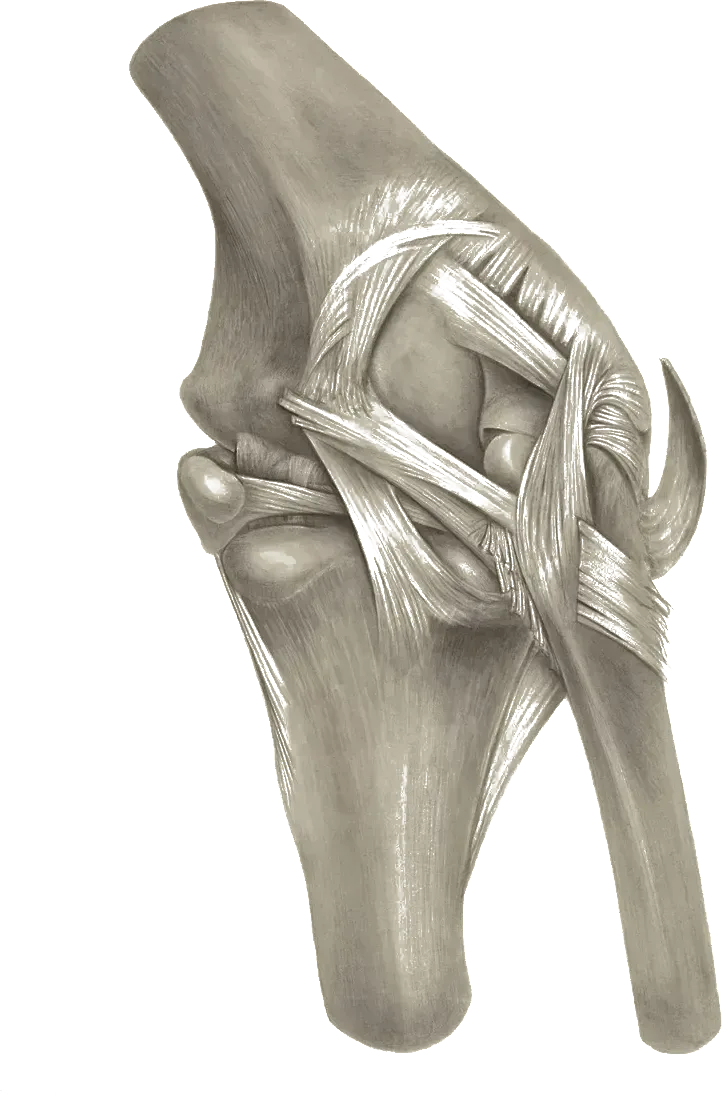
Link copiedThe (MCL) serves as the primary restraint against forces that push the knee into a knock-kneed position. This ligament has substantial tensile strength. The superficial MCL provides 57% of the restraining valgus moment at 5 degrees of knee flexion and increases to 78% at 25 degrees, showing that the ligament's contribution varies with knee angle. This angle-dependence explains why MCL injuries often occur with the knee in slight flexion rather than full extension.
MCL injuries occur far more frequently than LCL injuries due to the relative exposure to valgus-producing forces. When a football player gets struck on the lateral (outside) aspect of their knee, the impact creates a valgus force that stresses the MCL on the medial side. This mechanism accounts for the majority of MCL injuries in contact sports. The force required varies based on knee position, muscle activation, and impact direction, and a sufficiently large lateral blow can cause MCL failure when protective muscle activation is insufficient.
The lateral collateral ligament (LCL) resists forces that push the knee into a bow-legged position. This ligament has significantly lower tensile strength than the MCL. At 30 degrees of knee flexion, the LCL serves as the primary stabilizer against varus stress, bearing the majority of restraining forces. Pure varus-directed contact is relatively uncommon compared to valgus forces, explaining the lower incidence of isolated LCL injuries. When LCL injuries do occur, they often involve more complex mechanisms including hyperextension or rotational components.
The collateral ligaments work in concert with the and joint capsule to provide knee stability. When your MCL is intact, it prevents the medial joint space from gapping open during valgus stress. If the MCL tears completely, the tibia can translate laterally relative to the femur, creating joint . Research using instrumented knee testing shows that isolated MCL tears allow increased medial joint opening at 30 degrees flexion, while combined MCL and posterior oblique ligament tears produce still greater opening, demonstrating the importance of secondary restraints in maintaining stability.
Athletic movements involving cutting and pivoting create dynamic valgus loads on the MCL. When you plant your foot and cut to change direction, your body's momentum creates an external rotation and valgus moment at the knee. If your quadriceps, hamstrings, and hip muscles don't activate quickly enough to control this motion, excessive valgus stress loads the MCL. Studies using motion capture in soccer players show that poorly controlled cutting maneuvers generate increased knee valgus angles, creating MCL forces approaching injury thresholds. Athletes with weak hip demonstrate greater dynamic valgus during cutting, increasing MCL injury risk.
Ski injuries create unique mechanisms for both MCL and LCL damage. The MCL "phantom foot" injury occurs when a skier catches their inside edge and falls backward with the knee twisted inward, creating combined valgus and external rotation forces. The ski boot acts as a long lever arm, amplifying forces transmitted to the knee. Biomechanical analysis of ski falls shows that the rigid boot substantially increases valgus moments compared to athletic shoes, explaining why MCL tears are among the most common knee injuries in recreational skiing.
Previous MCL injuries alter knee even after clinical healing. After an MCL sprain, the ligament often heals with increased length (), reducing its ability to restrain valgus forces. Some athletes develop chronic medial knee laxity after MCL injuries, demonstrating greater joint opening compared to the uninjured side during valgus stress testing. This residual laxity increases the risk of future MCL injuries and can contribute to premature medial compartment knee due to altered loading patterns.
Contact sports create the highest risk for MCL injuries through direct trauma mechanisms. In football, rugby, hockey, and soccer, lateral knee contact during tackling or blocking generates high-magnitude valgus forces. Tackle impacts can generate high peak forces capable of exceeding the MCL's failure threshold. The combination of high force magnitude, rapid loading rate, and inability to generate protective muscle contraction before impact makes these injuries particularly common and often severe in contact athletes.
Symptoms
Clinical Presentation
Link copiedPrimary Symptoms
Associated Symptoms
Typical pattern
Acute injury with specific mechanism. Pain and swelling localized to ligament.
Symptoms
Differential Diagnosis
Link copiedConditions with similar presentations:
Medial Meniscus Tear
Key differences: Joint-line tenderness rather than tenderness along the course, mechanical catching or locking, positive McMurray or Thessaly test. Often follows a twisting mechanism with the foot planted rather than a pure blow.
ACL Tear with Associated MCL Injury
Key differences: Large acute effusion within hours, pop at time of injury, positive Lachman and pivot shift, and a sense of the knee giving way. An isolated sprain typically produces more localized swelling over the ligament and a stable Lachman.
Posterolateral Corner Injury
Key differences: , increased external rotation at 30 degrees on the dial test, and often a hyperextension or posterolateral blow mechanism. Easy to miss when attention stays fixed on the obvious lateral-sided pain and commonly co-occurs with injuries.
Pes Anserine Bursitis or Tendinopathy
Key differences: Pain two to three fingerbreadths below the medial joint line at the pes insertion rather than over the proper, gradual onset with overuse rather than a distinct trauma, and no on stress testing.
Iliotibial Band Syndrome
Key differences: Lateral knee pain at the that can be confused with pain, but it is reproduced by repetitive knee flexion-extension such as running rather than by stress, and there is no on clinical testing.
Tibial Plateau or Proximal Fibula Fracture
Key differences: High-energy mechanism, bony rather than ligamentous tenderness, and often inability to bear weight. Proximal fibula tenderness after a injury in particular should raise concern for an associated with or posterolateral corner disruption and warrants imaging.
When to seek professional help
Research
Key Research & Evidence
Peer-reviewed studies supporting the treatment approach.
Finding
Non-operative rehabilitation is promising for isolated MCL injuries, but evidence certainty is low
Research details
A 2024 systematic review by Svantesson and colleagues, titled 'Shedding light on the non-operative treatment of the forgotten side of the knee: rehabilitation of medial collateral ligament injuries' (BMJ Open Sport & Exercise Medicine), concluded that non-operative rehabilitation of MCL injuries appears promising, while cautioning that the supporting evidence is of very low certainty with substantial heterogeneity and no clear difference in outcomes between grade I and grade II injuries
Clinical relevance
Evidence supports conservative physiotherapy as primary treatment even for complete MCL tears when ligament remains in anatomical position and no associated injuries present
Finding
Weak hip abductors are associated with greater dynamic knee valgus and increased injury risk
Research details
Motion-analysis research on cutting and pivoting indicates that reduced hip abductor strength and control are associated with greater dynamic knee valgus, which increases medial-sided loading and is recognised as a modifiable contributor to collateral ligament injury risk
Clinical relevance
Hip strengthening forms critical component of MCL rehabilitation and injury prevention programs to address proximal control deficits contributing to knee valgus loading
Research Database Expanding
Additional peer-reviewed studies are being reviewed and will be added to strengthen the evidence base for this condition.
Management
Evidence-Based Management
Treatment strategies with the strongest support in the current literature.
Primary approach
Most Grade I and II /LCL sprains return to sport with structured conservative care built around early mobilization and progressive strengthening
Complementary
Functional bracing provides stability during healing phases while allowing controlled movement and preventing joint stiffness
Prevention & long-term
Neuromuscular training programs focusing on landing mechanics and knee control can meaningfully reduce injury risk in pivoting sports
Detailed management strategies
Bracing if Needed
Provides stability during healing
Important precautions
- Don't become dependent on brace
Progressive Loading
Stimulates ligament healing
Important precautions
- Respect healing timeframes
Management
Treatment Techniques
Evidence-based manual therapy and intervention approaches.
Treatment approaches supported by current research and clinical guidelines
Recommended treatment approaches
Treatment approaches are individualized to each patient's needs and goals. All interventions require explicit informed consent, and treatment plans are collaboratively modified based on your preferences and response to care.
Sports Rehabilitation & Return to Sport
Evidence-based recovery programs for athletes to safely return to sport after injury.
Post-Surgical Rehabilitation
Evidence-based recovery programs following surgery to restore function and strength.
Rehabilitation
A Typical Rehabilitation Progression
Three phases, from settling symptoms to returning to full activity.
Recovery from MCL is usually staged: calm the symptoms first, then rebuild the strength and capacity of the area, then return to your full activities. The three phases below show the kind of progression the evidence supports and that I commonly work through in clinic. They are here to show you what the road can look like, not to act as a personal program.
- Phase 1
Protected Healing (Weeks 0 to 3)
Control swelling, protect the ligament from further or stress, restore range of motion within comfortable limits, and keep the quadriceps active. The Logerstedt et al. JOSPT CPG on knee stability and movement coordination impairments supports early protected motion over immobilization for injuries, because prolonged rest weakens both the ligament and the surrounding musculature.
Examples, not a prescription
- Quad sets and straight leg raises in all four planes to maintain quadriceps volume without valgus or varus load
- Pain-free active and active-assisted knee flexion and extension, progressing range as guarding settles
- Stationary bike once flexion reaches approximately 100 degrees, with a high seat and low resistance
- Hinged knee brace for Grade II and III injuries during walking and light daily activity
- and external rotator isometrics in standing or sidelying to preload proximal control
Ready to progress when
Minimal to trace effusion, near-symmetrical range of motion, ability to walk without a limp inside the brace, quadriceps activation with no extension lag on , and pain-free low-load closed-chain work.
- Phase 2
Strength and Single-Leg Control (Weeks 3 to 8)
Rebuild bilateral and single-leg strength, reintroduce frontal-plane loading under control, and restore confidence in valgus and varus directions. This is the phase where I see the most avoidable setbacks, because patients feel good and skip the slow work that actually protects the ligament.
Examples, not a prescription
- Progressive leg press, split squats, and Romanian deadlifts building toward limb symmetry above 80 percent
- Step-ups and step-downs with a mirror or video check to catch knee collapse into valgus
- Lateral band walks, single-leg hip thrusts, and Copenhagen holds to load the medial chain under control
- Single-leg balance progressions on firm then unstable surfaces, adding head and trunk movements
- Pool running and gradual on-land jog-walk intervals once straight-line running is tolerated without reactive swelling
Ready to progress when
Quadriceps and hamstring limb symmetry around 85 to 90 percent, pain-free single-leg squat with controlled frontal-plane alignment, and effusion that does not flare within 24 hours of training.
- Phase 3
Return to Cutting and Sport (Weeks 6 to 12+, grade-dependent)
Reintroduce change-of-direction, deceleration, and contact tolerance. Grade I injuries often reach this phase around weeks 4 to 6. Grade III isolated MCL injuries more typically enter it at 8 to 12 weeks. The point of the phase is not calendar time, it is demonstrating that the knee tolerates the specific demands of the sport being returned to.
Examples, not a prescription
- Hop test battery including single hop for distance, triple hop, crossover hop, and timed 6 metre hop targeting limb symmetry of 90 percent or greater
- Progressive change-of-direction work: 45 degree cuts, 90 degree cuts, then reactive cutting off a partner or cue
- progression from bilateral to unilateral landings, adding deceleration and lateral bounding
- Sport-specific drills that reintroduce planned and unplanned valgus or varus loading under supervision
- Return-to-contact progression for collision sports, starting with controlled drills before full competition
Ready to progress when
Limb symmetry index of 90 percent or greater on strength and hop testing, clean change-of-direction mechanics without valgus collapse, no effusion response to training, and subjective confidence in the knee during sport-specific tasks.
Management
Prognosis & Recovery
What outcomes and recovery factors typically look like.
Expected timeline
Grade I: 2-4 weeks, Grade II: 4-8 weeks, Grade III: 8-12 weeks
Natural history
Good healing potential with appropriate management
Factors affecting recovery
Management
Measuring Progress
How to track the recovery arc week to week.
Day-to-day tracking
I track what changes day to day: pain interference with key tasks, movement quality during functional tests, and your confidence with daily activities
Assessment tools
Condition-specific questionnaires when useful (like the Oswestry for back pain or DASH for shoulder conditions)
Activity targets
One activity target that matches your goal - whether that's returning to sport, work tasks, or daily activities without limitation
Management
Frequently Asked Questions
Common concerns and answers about this condition.
Do I need surgery for a torn MCL?
Do I need surgery for a torn MCL?
Usually not. Isolated tears, including complete Grade III injuries, heal well with structured rehabilitation in the large majority of cases. This is one of the few knee ligaments with strong biological healing capacity, and older natural-history work by Indelicato and more recent systematic reviews consistently support non-operative management as the default. Surgery is considered when the MCL is torn in combination with an , PCL, or posterolateral corner injury, when the tibial-sided fails to heal, or when persistent remains after a full rehabilitation trial.
Is LCL injury treated the same way as MCL?
Is LCL injury treated the same way as MCL?
No, and this is where I try to be honest with patients. The is a broad ligament with good blood supply and heals predictably. The LCL is a smaller cord-like structure with less favourable healing, and it rarely injures in isolation. Grade I and II LCL sprains can be managed conservatively, but a Grade III LCL tear, especially with any posterolateral corner involvement, often needs a surgical opinion early. Missing a posterolateral corner injury and rehabbing it as a simple sprain is a known way to end up with a chronically unstable knee.
How long until I can run and return to sport?
How long until I can run and return to sport?
Grade I sprains tend to return to running in 2 to 3 weeks and sport by 4 weeks. Grade II is typically 4 to 8 weeks, with sport-specific cutting work starting once single-leg strength and control approach the other side. Grade III isolated injuries often take 8 to 12 weeks for return to sport when the rest of the knee is stable. These are ranges, not promises. I use criteria like pain-free single-leg squat, symmetrical hop performance, and confidence on cutting drills rather than a pure calendar date.
Do I need to wear a knee brace?
Do I need to wear a knee brace?
A hinged knee brace limiting or stress is reasonable during the early healing phase for Grade II and III injuries, and I often use one to let patients stay mobile without worrying about an awkward step. It is not a lifetime commitment. The goal is to wean off the brace as strength and neuromuscular control return, because long-term bracing does not strengthen the ligament and can create a dependency that slows confidence-building.
Will I always be unstable after an MCL tear?
Will I always be unstable after an MCL tear?
Most people regain functional stability, but some end up with a small amount of residual on clinical testing that never fully disappears. What matters more is whether the knee feels stable during cutting, pivoting, and deceleration, and that is driven by quadriceps strength, hip control, and neuromuscular retraining rather than by millimetres of laxity on an exam table. Patients who commit to progressive strengthening and return-to-sport testing overwhelmingly report a knee they trust, even if the ligament itself heals slightly longer than the other side.
Do I need an MRI?
Do I need an MRI?
For a clear isolated sprain with a typical mechanism, localized medial tenderness, and stable , Lachman, and posterior drawer testing, imaging is not mandatory to start rehabilitation. I send for MRI when the mechanism or exam suggests a combined injury, when the LCL or posterolateral corner is in question, when there is joint-line tenderness suggesting a tear, or when a patient is not progressing as expected. Imaging should change the plan, not just reassure.
Can I still exercise during recovery?
Can I still exercise during recovery?
Yes, and I strongly prefer that you do. Early protected loading and range-of-motion work produce better outcomes than immobilization, which is supported by the rehabilitation literature. Upper body training, stationary cycling within comfortable range, hip and core work, and single-leg balance on the uninjured side can all continue. What changes is the dose and the knee-specific exercises, not the fact of training.
Will this cause arthritis later?
Will this cause arthritis later?
Isolated injuries that heal well do not have a strong link to later . The risk rises when the MCL injury is part of a multi-ligament event, when there is an associated tear, or when the knee is left with persistent and altered loading. That is part of why I do not treat a grade III MCL as a trivial injury, even when it heals without surgery: the quality of the rehabilitation affects the mechanical environment of the joint for years afterward.
Related Conditions
Conditions I commonly see alongside, or confused with, this one.
- Anatomically related
ACL Injuries
Multi-ligament knee injuries common; often occur together in trauma
- Anatomically related
Meniscal Injuries
Complex knee injuries often involve both ligaments and meniscus
- Common co-occurrence
Osteoarthritis of the Knee
Ligament injuries can lead to joint instability and secondary arthritis