Hip Osteoarthritis
Joint degeneration, cartilage breakdown, activity-related pain
Overview
The Science of Hip Osteoarthritis
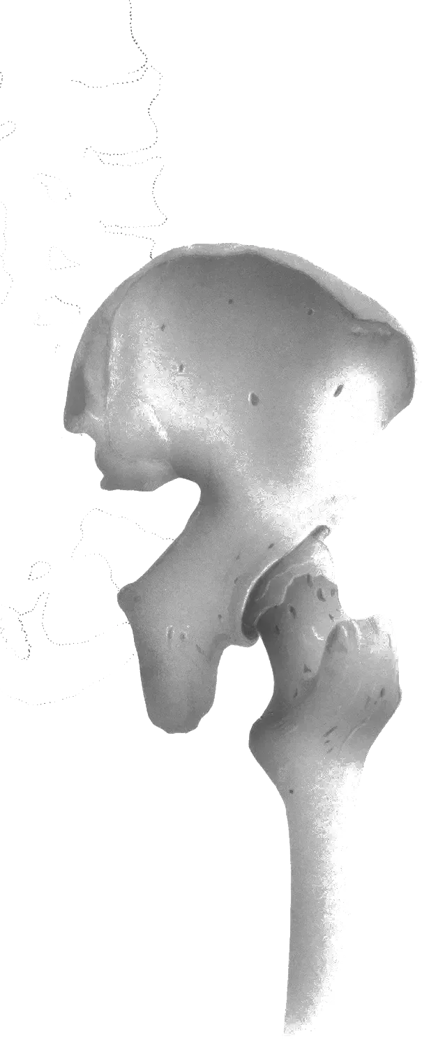
Link copiedHip is a dynamic process involving the entire joint structure, not simple "wear and tear." The story of hip osteoarthritis rarely begins with a bang - it's a slow burn that often starts as a subtle, deep, groin-area ache noticed after a long walk that might be dismissed as a simple muscle strain. The most pervasive and damaging misconception about OA is that it's a simple "wear and tear" disease where the joint is worn out and nothing can be done. This is not the full picture. OA is a dynamic process involving the entire joint structure, and it does not mean your active life is over. It begins with articular cartilage breakdown (the smooth, white, slippery tissue covering bone ends), followed by subchondral bone changes, formation ("bone spurs" - bony lumps growing in response to inflammation), and joint space narrowing. The process involves inflammatory mediators, altered , and compensatory muscle weakness. Critically, pain is not directly proportional to radiographic changes - I regularly see patients with "severe" OA on imaging who have minimal pain and excellent function, and vice versa. Your experience of pain is real, but it is not solely dictated by what an X-ray shows.
The hip joint doesn't exist in isolation - when it becomes stiff and painful from OA, the body makes compensations. The most common is increased movement and strain on the and (SI) joint, which is why so many people with hip OA also develop low back pain. The knee can also take a hit - a stiff hip changes the way you walk, altering forces that travel down through the knee and ankle. Living with persistent pain is exhausting and can lead to fear of movement, anxiety, and feeling of fragility. This is where pain comes in - over time, the nervous system can become , essentially "turning up the volume" on pain signals. Gentle, graded movement can help recalibrate the nervous system. Hip osteoarthritis may coexist with other hip conditions such as greater pain syndrome or hip , and can develop secondary to previous conditions like femoroacetabular (FAI) or hip tears.
Overview
Contributing Factors
Link copiedYour hip joint bears substantial forces during daily activities - approximately 2.4 times your body weight during normal walking, increasing to 2-3 times body weight with faster walking. This load multiplication explains why even modest weight gain significantly impacts hip joint stress. Research using instrumented hip implants shows that walking at approximately 4 km/h generates peak forces of 238% body weight, with heel strike creating the highest loading moments.
In hip , the biomechanical picture changes substantially. Recent systematic reviews (2023-2024) reveal that individuals with mild-to-moderate hip OA experience less net hip joint loading over a reduced range of hip motion for a longer proportion of the gait cycle. This means you're spreading lower forces over longer periods through less movement - a compensatory strategy that reduces instantaneous peak loads but perpetuates stiffness and muscle weakness.
The altered gait mechanics create a problematic cascade: reduced hip motion forces adjacent joints (your lower back and ) to compensate by moving more, while your knee experiences altered force distribution. Research specifically shows loading alterations in adjacent and contralateral joints in knee OA but interestingly not in hip OA to the same degree, though the reduced hip motion itself becomes the primary biomechanical driver of dysfunction.
Muscle function plays a critical role. The hip (particularly and minimus) normally stabilize your pelvis during single-leg stance. When these weaken in OA, you develop a pattern - your pelvis drops on the unsupported side during walking, which increases compressive forces on the already compromised joint. This muscle weakness isn't just a consequence of pain avoidance; studies show actual neuromotor changes including muscle co-contraction patterns that increase joint loading while paradoxically reducing effective force production.
Modern research (2024) emphasizes that discrepancies exist between external joint moments (what we measure) and internal joint loads (what your cartilage experiences) in people with OA due to altered neural patterns and muscle co-contraction. This explains why two people with identical radiographic OA can have completely different pain and function levels - the biomechanical loading patterns differ based on neuromuscular control strategies.
Symptoms
Clinical Presentation
Link copiedPrimary Symptoms
Associated Symptoms
Typical pattern
The story rarely begins with a bang - it's a slow burn. Often starts as a subtle, deep, groin-area ache noticed after a long walk that might be dismissed as a muscle strain. Then, you start to feel profound stiffness first thing in the morning, making it a real chore to put on your socks and shoes. Over months or even years, that ache becomes more persistent, and the stiffness takes longer to fade. The narrative I hear from patients is consistent: a gradual closing down of their world. Activities they once loved, like hiking, gardening, or playing with grandchildren, are now shadowed by the thought of the pain that might follow. Classic presentation: age >40, activity-related groin pain, morning stiffness that improves with gentle movement, limited internal rotation on examination.
Symptoms
Differential Diagnosis
Link copiedConditions with similar presentations:
Greater Trochanteric Pain Syndrome (Gluteal Tendinopathy)
Key differences: Point tenderness on the outer hip over the rather than groin pain, severe pain lying on the affected side at night, and a preserved passive hip internal rotation compared with hip .
Femoroacetabular Impingement Syndrome
Key differences: Sharp anterior groin pain with deep flexion and internal rotation in a younger active patient, positive , and mechanical catching during squats or getting out of a low car. Morning stiffness is usually brief compared with .
Lumbar Spine Referral (Facet or Radicular Pain)
Key differences: Pain changes with movement rather than hip movement, hip passive range is full and symmetrical, and pain may travel below the knee. Hip rotation is preserved on the examination table.
Inflammatory Arthropathy (Ankylosing Spondylitis or Rheumatoid)
Key differences: Morning stiffness longer than an hour that does not settle with movement, systemic symptoms such as fatigue or rashes, bilateral involvement, and inflammatory markers on bloodwork.
Hip Labral Tear
Key differences: Clicking or catching with pivoting, a from groin to lateral hip, and mechanical symptoms more prominent than morning stiffness. Often coexists with FAI morphology in younger patients.
Stress Fracture of the Femoral Neck
Key differences: Groin pain that worsens rather than improves with activity, significant pain with hopping or single-leg loading, history of a recent spike in running or military-style training, and red flags in the hormonal or nutritional history.
When to seek professional help
Research
Key Research & Evidence
Peer-reviewed studies supporting the treatment approach.
Study
Hip Pain and Mobility Deficits - Hip Osteoarthritis: Revision 2017 (JOSPT Clinical Practice Guidelines)
Key findings
Strong recommendation for individualised exercise therapy and for manual therapy combined with exercise to improve pain, mobility, and function in hip osteoarthritis
Clinical relevance
Establishes conservative care as evidence-based first-line treatment
Study
Total Hip Replacement or Resistance Training for Severe Hip Osteoarthritis (PROHIP)
Key findings
In severe hip osteoarthritis, total hip replacement led to greater improvement in self-reported pain and function at 6 months than a resistance training programme (mean Oxford Hip Score change about 15.9 vs 4.5 points)
Clinical relevance
Clarifies that surgery can outperform exercise in severe disease, while conservative care remains a reasonable first-line option in milder presentations
Research Database Expanding
Additional peer-reviewed studies are being reviewed and will be added to strengthen the evidence base for this condition.
Management
Evidence-Based Management
Treatment strategies with the strongest support in the current literature.
Primary approach
Exercise therapy can match surgery for pain and function in many patients, with most seeing meaningful improvement over several months
Complementary
Patient education about load-capacity principles and activity modification enables self-management and reduces fear-avoidance behaviors that contribute to disability
Prevention & long-term
Early intervention with strengthening and mobility exercises can delay progression and, for some patients, postpone joint replacement
Detailed management strategies
Load vs Capacity Balance
Pain flares when demands exceed hip's current capacity. Temporary reduction of aggravating loads while building capacity through exercise
Important precautions
- Avoid complete rest
- Gentle movement helps morning stiffness
- Monitor symptom response to activities
Progressive Exercise Program
Strong muscles act as shock absorbers, reducing stress on cartilage. Exercise stimulates cartilage health
Important precautions
- Start with pain-free range
- Progress gradually
- Consistency more important than intensity
Heat for Stiffness
Heat therapy helpful for morning stiffness and before exercise
Important precautions
- Use before activity, not for acute inflammation
Management
Treatment Techniques
Evidence-based manual therapy and intervention approaches.
Treatment approaches supported by current research and clinical guidelines
Recommended treatment approaches
Treatment approaches are individualized to each patient's needs and goals. All interventions require explicit informed consent, and treatment plans are collaboratively modified based on your preferences and response to care.
Pain Education & Self-Management
Understanding pain science to reduce fear and improve movement confidence alongside active rehabilitation.
Exercise Therapy
Personalized exercise programs designed to restore strength, flexibility, and function.
Joint Mobilization
Graded techniques to restore joint movement and reduce stiffness.
Rehabilitation
A Typical Rehabilitation Progression
Three phases, from settling symptoms to returning to full activity.
Recovery from Hip Osteoarthritis is usually staged: calm the symptoms first, then rebuild the strength and capacity of the area, then return to your full activities. The three phases below show the kind of progression the evidence supports and that I commonly work through in clinic. They are here to show you what the road can look like, not to act as a personal program.
- Phase 1
Calming the Joint and Restoring Tolerance (Weeks 1 to 4)
The 2025 JOSPT Hip CPG revision (Koc, Cibulka et al., doi:10.2519/jospt.2025.0301) gives a strong recommendation for patient education and individualised exercise as first-line care. Early weeks are about removing friction, not chasing strength. I find my tolerable dose of walking, standing, and stair work, introduce low-compression movement to lubricate the joint, and pair it with a short daily warm-up for morning stiffness.
Examples, not a prescription
- Supine hip internal and external rotation through comfortable range, 2 sets of 10 slow repetitions, morning and evening to shorten start-up stiffness
- Stationary bike with a high seat and easy resistance, 8 to 12 minutes, which the Cochrane review of exercise for hip OA (Fransen et al., 2014) supports as a reliable pain-reducing modality
- Supine or seated pressing against a belt or wall, 4 sets of 30 seconds, to begin reloading the abductors without joint flare
- Walking in tolerable bouts split across the day rather than one long walk, scaled to end below a 3 out of 10 ache
- Heat before movement, short rotation circles before getting out of bed, and pacing cues for stairs and car entry
Ready to progress when
Morning stiffness shortened to under 20 minutes on most days, a 20 to 30 minute continuous walk managed without a next-day flare, and the patient can explain the load-capacity model in their own words.
- Phase 2
Strengthening the Hip Envelope (Weeks 5 to 12)
This mirrors the structured land-based exercise recommended by OARSI 2019 (Bannuru et al., Osteoarthritis and Cartilage) as a Core Treatment for hip OA. The goal here is building a strong hip envelope, particularly abductors and extensors, so the joint is shared-loaded across muscle rather than dumped into bone. Resistance is progressed weekly by about 10 percent when the 24-hour response stays quiet.
Examples, not a prescription
- Sit-to-stand from a chair at a height that allows pain below 3 out of 10, 3 sets of 8 to 12 reps, progressing to a lower chair across the block
- Hip hinge to a chair or bench with bodyweight progressing to a goblet hold, 3 sets of 10, teaching a posterior chain strategy for bending and lifting
- Supported split squat with hands on a bench, shallow depth at first, 3 sets of 8 per side
- Banded standing hip abduction with a cue to keep the pelvis level, 3 sets of 12 per side
- Continuous stationary bike or pool walking 20 to 30 minutes, 3 times weekly, for aerobic capacity and joint lubrication
Ready to progress when
Can rise from a standard dining chair without pushing off the thighs, walks 30 to 45 continuous minutes without next-day flare, and tolerates 3 sets of 10 hip hinges with an 8 to 12 kilogram load.
- Phase 3
Loading for Real-World Activity (Months 3 to 6 and beyond)
At this point the hip tolerates moderate loads and the patient wants their activities back, whether that is hiking, gardening for an afternoon, playing with grandchildren, or getting back to yoga or pickleball. Loading is progressed into single-leg work, hills, and patient-specific demands. This aligns with the 2025 JOSPT CPG's functional, gait, and balance training recommendations.
Examples, not a prescription
- Step-ups to a 15 to 20 centimetre step with a focus on driving through the whole foot, 3 sets of 8 per side
- Goblet squat to a box, gradually lowering the box across the block as range and confidence improve, 3 sets of 8
- Single-leg Romanian deadlift to a light touch of a chair or wall, 3 sets of 6 per side, for posterior chain control
- Graded hill walking or loaded carries for patients whose goals include hiking or travel with luggage
- Return to chosen recreation with a week-by-week exposure plan rather than an all-or-nothing comeback
Ready to progress when
Sustained return to the activities the patient cared about at intake, confidence on stairs and single-leg tasks, morning stiffness brief or absent, and a maintenance programme the patient is actually willing to do twice a week.
Management
Prognosis & Recovery
What outcomes and recovery factors typically look like.
Expected timeline
Managing a chronic condition like is a long-term project, not a quick fix. You can expect to see meaningful improvements in pain and function within 6-12 weeks of consistent effort, but the true benefits come from integrating these strategies into your lifestyle permanently. There will be good days and bad days. Realistic markers of progress include: Can you walk for 5 minutes longer than last month? Do you feel more confident going up and down stairs? Is the morning stiffness lasting 15 minutes instead of 30? Did you get back to gardening for an hour without a major flare-up?
Natural history
Progressive structural changes, but symptoms fluctuate significantly. Many patients maintain excellent function with proper management. The key insight is that pain is not directly related to imaging findings - your experience is real but not determined by X-ray appearance. Living with persistent pain can lead to fear of movement, anxiety, and feeling of fragility, but gentle, graded movement can help recalibrate the nervous system.
Factors affecting recovery
Management
Measuring Progress
How to track the recovery arc week to week.
Day-to-day tracking
I track what changes day to day: pain interference with key tasks, movement quality during functional tests, and your confidence with daily activities
Assessment tools
Condition-specific questionnaires when useful (like the Oswestry for back pain or DASH for shoulder conditions)
Activity targets
One activity target that matches your goal - whether that's returning to sport, work tasks, or daily activities without limitation
Management
Frequently Asked Questions
Common concerns and answers about this condition.
Do I need an X-ray before starting physiotherapy for hip osteoarthritis?
Do I need an X-ray before starting physiotherapy for hip osteoarthritis?
Usually no. The 2025 JOSPT Hip Clinical Practice Guideline and OARSI 2019 both describe hip OA as a clinical diagnosis based on age, symptom pattern, and examination findings. An X-ray can confirm the picture when it is unclear or when surgery is being considered, but X-ray severity correlates poorly with pain and function. I have seen patients with minimal changes on imaging who are in significant pain, and others with severe changes who walk for an hour without symptoms.
Will exercise make my hip arthritis worse?
Will exercise make my hip arthritis worse?
No. The Cochrane review of exercise for hip (Fransen et al., 2014) showed reliable small-to-moderate reductions in pain and improvements in function, and no signal of harm. The 2025 JOSPT CPG strongly recommends individualised strengthening, flexibility, and endurance programmes. What makes hip OA worse is a sudden spike beyond current capacity, not loading itself. Dosing matters more than avoidance.
Can physiotherapy help me avoid a hip replacement?
Can physiotherapy help me avoid a hip replacement?
For many people, yes, at least for years. Strong exercise evidence backs meaningful symptom and function improvement. That said, I will not oversell it. The Bennell JAMA 2014 trial specifically found that a structured multimodal physiotherapy programme did not outperform sham over 36 weeks, which has been debated widely since. My read of the broader literature, including the 2025 JOSPT revision, is that consistent long-term loading, weight management, and education meaningfully shift the trajectory for most patients, and surgery remains a reasonable option when conservative care has plateaued and pain is dominating daily life.
Why does my hip hurt in the groin rather than the side?
Why does my hip hurt in the groin rather than the side?
The hip joint sits deeper and more anterior than most people expect, so genuine hip joint pain referred from articular cartilage and the is most often felt in the groin and front of the thigh, occasionally travelling to the knee. Pain over the outer hip bone usually points to the gluteal tendons rather than the joint itself. This is why I examine hip rotation carefully. Losing internal rotation first is one of the most reliable examination findings in hip .
Is it safe to keep running with hip osteoarthritis?
Is it safe to keep running with hip osteoarthritis?
In many cases, yes, with adjustments. Running is not the cause of hip in most people. Total volume, terrain, recovery, and overall hip strength matter more than the act of running itself. I usually keep symptomatic runners running at a reduced, symptom-guided dose while they build and extensor capacity, then rebuild volume by about 10 percent per week. Trail running and downhill running are the two patterns I temporarily reduce first.
What about glucosamine, collagen, and supplements?
What about glucosamine, collagen, and supplements?
OARSI 2019 issued conditional recommendations against glucosamine and chondroitin for hip , citing inconsistent evidence on pain and function. and turmeric data are mixed. I do not discourage patients from trying low-risk options, but I do not want supplements displacing the interventions with the strongest evidence, which are exercise, education, weight management where relevant, and topical or short-course oral NSAIDs on flare days under their physician's guidance.
How long until I feel a real difference?
How long until I feel a real difference?
Most patients notice meaningful change in morning stiffness and walking tolerance within 4 to 6 weeks of consistent loading. Strength adaptations and larger functional gains land closer to the 3-month mark. The 2025 JOSPT CPG and OARSI 2019 both describe hip management as an ongoing project rather than a short course of treatment. My goal is to get you to a programme you will actually keep doing twice a week for the long term.
Does cracking or grinding in the hip mean it is getting worse?
Does cracking or grinding in the hip mean it is getting worse?
No. is common and does not correlate well with joint damage or outcomes. A quiet hip is not necessarily a healthier hip. If clicking comes with sharp pain, giving way, or true mechanical locking, that changes the conversation and I examine for involvement. Painless noise on its own is not a reason to stop exercise.
Related Conditions
Conditions I commonly see alongside, or confused with, this one.
- Biomechanically linked
Osteoarthritis of the Knee
Hip stiffness alters gait and can increase knee joint stress leading to arthritis
- Common co-occurrence
Femoroacetabular Impingement (FAI)
FAI can lead to cartilage damage and secondary osteoarthritis
- Anatomically related
Lateral Hip Pain & Gluteal Tendinopathy
Both affect hip region; GTPS often coexists with hip OA
Commonly confused with
Side-by-side comparisons for patterns that often get mistaken for hip osteoarthritis.