Facet Joint Syndrome
Facet joint pain and dysfunction
Overview
The Science of Facet Joint Syndrome
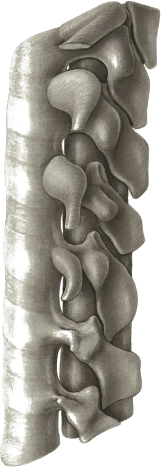
Link copiedare small joints between vertebrae that guide and limit movement. They can become arthritic or inflamed, causing localized back pain. The joint capsule is richly innervated, making it a significant pain source.
Overview
Contributing Factors
Link copiedThe function as part of a three-joint complex at each spinal level, working together with the intervertebral disc anteriorly and two facet joints posteriorly. This mechanical relationship means that changes in any one component directly affect loading patterns on the others. When your intervertebral disc degenerates and loses height, the fibers shift loads more posteriorly onto the facet joints, accelerating their process. Finite element modeling shows that disc degeneration shifts load posteriorly and substantially increases facet joint loading compared to healthy discs.
During spinal extension (leaning backward), the inferior articular process of the upper vertebra moves inferiorly and posteriorly until the spinous processes approach contact. In this position, facet joint loading increases substantially compared to neutral spine alignment. Biomechanical studies using pressure-sensitive film inserted into cadaveric facet joints demonstrate that extension movements markedly increase facet joint contact forces relative to neutral standing posture. This explains why activities involving repeated or sustained extension, such as overhead work, backward bending, or arching movements, frequently trigger facet joint pain.
The lumbar extensor muscles amplify facet joint compression during active extension movements. When you actively arch your back, your erector spinae and muscles contract to produce the movement, but these same muscles generate compressive forces that push the vertebrae together. This muscle-generated compression adds to the forces already present from body weight and spinal positioning. Active extension movements create higher facet loading than passive extension to the same position, which can speed degenerative changes over time.
Combined extension with rotation movements create particularly high facet joint stresses. When you twist your spine while bending backward, one facet joint experiences increased compression while the opposite side experiences tensile and shear forces. This asymmetrical loading pattern concentrates stress on specific portions of the joint surfaces. Studies on golfers, who repeatedly combine extension and rotation during their swing, show accelerated unilateral facet joint degeneration on the lead side (left side for right-handed golfers), with degeneration patterns correlating directly with swing .
Asymmetric facet joint degeneration creates altered spinal mechanics that perpetuate the problem. Recent finite element analysis research (2025) investigating asymmetric L4-L5 facet degeneration demonstrates that when one facet joint degenerates more than its paired counterpart, it alters the entire segment's movement behavior. The degenerated joint experiences higher contact forces and increased range of motion, while the opposite joint may become stiffer. This mechanical imbalance creates a self-perpetuating cycle where asymmetric wear patterns progressively worsen.
Repetitive loading during occupational and recreational activities accumulates facet joint stress over time. Jobs requiring prolonged standing, frequent overhead reaching, or repetitive backward bending expose facet joints to thousands of high-load cycles daily. Physically demanding occupations with sustained overhead reaching and repetitive backward bending are associated with higher rates of facet joint degeneration than low-demand work. The cumulative nature of this mechanical stress means that even moderate loads, when repeated frequently, can exceed the joint's capacity for repair and adaptation.
Spinal and facet joint hypertrophy create a biomechanical feedback loop. As facet joints degenerate, they often develop (bone spurs) and thickened joint capsules in response to abnormal mechanical stress. This hypertrophy can narrow the spinal canal and foramen, creating stenotic conditions. The stenosis then alters load distribution across the spine, potentially increasing stress on adjacent facet joints and propagating degenerative changes to multiple spinal levels. Studies tracking stenosis progression show that once this cascade begins at one level, adjacent levels develop stenotic changes at accelerated rates.
The facet joints' orientation in the lumbar spine makes them particularly vulnerable to extension and rotation forces. At L4-L5 and L5-S1, the facet joints sit more vertically oriented, while at upper lumbar levels they orient more horizontally. This anatomical variation means that lower lumbar facets resist more anterior shear forces, while upper lumbar facets resist more rotation. Transition zones where facet orientation changes, such as the thoracolumbar and lumbosacral junctions, are recognised sites of mechanical stress concentration during combined movements, and lower lumbar levels show the highest facet degeneration prevalence.
Symptoms
Clinical Presentation
Link copiedPrimary Symptoms
Associated Symptoms
Typical pattern
Worse with extension and rotation. Better with flexion. Morning stiffness that improves with movement.
Symptoms
Differential Diagnosis
Link copiedConditions with similar presentations:
Lumbar Discogenic Pain
Key differences: Central or slightly off-centre low back pain worse with sustained flexion, sitting, and forward bending. Eased by extension and standing. Sneezing and coughing can spike symptoms. Referral pattern often follows a if the disc is affecting a .
Lumbar Spinal Stenosis
Key differences: Bilateral leg heaviness or cramping with walking and standing that eases rapidly with sitting or leaning forward. Typically older adults. Walking tolerance limited by leg symptoms more than by back pain itself.
Sacroiliac Joint Dysfunction
Key differences: Pain pinpointed at the PSIS just below the belt line, reproduced by a cluster of SI provocation tests. Referral rarely extends below the knee. Extension and rotation provocation is less consistent than at the facets.
Spondylolysis or Spondylolisthesis
Key differences: Often a history of adolescent-onset back pain in repetitive extension sports (gymnastics, fast bowling, diving). Pain reproduced by loaded single-leg extension (Stork test). Imaging confirms a pars defect or slip. Management shifts to relative extension offloading and graded loading rather than extension provocation.
Hip Osteoarthritis
Key differences: Groin or anterior thigh pain with reduced and painful hip internal rotation, worse with weight-bearing and hip-loaded activities. Spinal positions do not consistently change symptoms.
Inflammatory Back Pain (Axial Spondyloarthritis)
Key differences: Insidious onset before age 40, significant morning stiffness lasting more than 30 to 60 minutes, improvement with exercise but not with rest, and often night-time waking with pain. Requires medical referral rather than being managed as mechanical facet pain.
When to seek professional help
Research
Key Research & Evidence
Peer-reviewed studies supporting the treatment approach.
Finding
Facet joints cause 27-40% of chronic low back pain cases
Research details
Facet joint pain occurs in 27% to 40% of patients with low back pain but is often overlooked or misdiagnosed, with no clear correlation between clinical examination, radiological findings, and clinical presentation complicating diagnosis
Clinical relevance
High prevalence emphasizes importance of considering facet joint pathology in differential diagnosis for chronic low back pain, particularly when imaging doesn't correlate with presentation
Finding
Conservative management effective in majority of cases
Research details
Most episodes of low back pain respond well to brief rest, activity modification, and physical therapy, with approximately 50% of cases improving within 1-2 weeks and up to 90% showing resolution within 6-12 weeks
Clinical relevance
Supports conservative physiotherapy approach as first-line treatment before considering interventional procedures, with expectation that most patients improve without invasive intervention
Finding
Clinical examination guides treatment selection
Research details
2020 comprehensive guidelines indicate Level II evidence with strong strength of recommendation for physical examination and clinical assessment in selecting patients for facet joint interventions at least 3 months after onset and failure of conservative management
Clinical relevance
Emphasizes thorough clinical assessment to identify appropriate candidates for physiotherapy versus those requiring more advanced interventions after adequate conservative trial
Management
Evidence-Based Management
Treatment strategies with the strongest support in the current literature.
Primary approach
combined with targeted strengthening is the most consistently supported approach for facet-driven pain and usually produces meaningful change within a couple of months
Complementary
Movement restoration and spinal strengthening exercises address underlying movement dysfunctions while building resilience against future episodes
Prevention & long-term
Regular spine strengthening and movement education can substantially reduce the risk of recurrent back pain episodes by improving spinal stability and movement quality
Detailed management strategies
Movement Variety
Prevents prolonged loading of facets
Important precautions
- Avoid prolonged extension
Core Exercises
Supports spine and reduces facet loading
Important precautions
- Maintain neutral spine
Management
Treatment Techniques
Evidence-based manual therapy and intervention approaches.
Treatment approaches supported by current research and clinical guidelines
Recommended treatment approaches
Treatment approaches are individualized to each patient's needs and goals. All interventions require explicit informed consent, and treatment plans are collaboratively modified based on your preferences and response to care.
Rehabilitation
A Typical Rehabilitation Progression
Three phases, from settling symptoms to returning to full activity.
Recovery from Facet Joint Syndrome is usually staged: calm the symptoms first, then rebuild the strength and capacity of the area, then return to your full activities. The three phases below show the kind of progression the evidence supports and that I commonly work through in clinic. They are here to show you what the road can look like, not to act as a personal program.
- Phase 1
Foundation: Offload Extension, Restore Segmental Mobility
The first phase reduces sustained extension loading, desensitises the irritated facet segment, and restores basic flexion-based mobility. A short period of extension offloading is about creating room to rebuild capacity, not a long-term strategy of avoidance.
Examples, not a prescription
- Cat-camel and pelvic tilts on hands and knees, 10 slow controlled repetitions
- Supine single and double knee-to-chest, 30 seconds, 2 to 3 times
- Child's pose or prayer stretch for segmental flexion, 30 seconds, 2 to 3 repetitions
- Dead bug with floor contact, 2 sets of 6 to 8 per side, emphasising neutral spine
- Short frequent walking bouts of 5 to 10 minutes, breaking up prolonged standing
Ready to progress when
Resting pain 3/10 or less, tolerance of 30 to 45 minutes of standing with only mild symptom increase, and comfortable lumbar flexion to touch mid-shin or further.
- Phase 2
Progressive Loading: Hip-Driven Movement and Trunk Strength
Facet-loaded presentations often ride on top of weak hip extensors and poor hip hinge mechanics, which pushes extension and rotation demand into the lumbar segments. This phase shifts work back to the hips and builds the trunk strength needed to keep the spine in a more tolerant zone under load.
Examples, not a prescription
- Hip hinge progression: broomstick hinge, kettlebell deadlift, single-leg Romanian deadlift, 3 sets of 6 to 10
- Glute bridge and hip thrust, 3 sets of 8 to 12
- Goblet squat to a box, 3 sets of 8 to 10, progressed in depth and load
- Side plank and Pallof press for lateral and anti-rotation trunk control
- Bird dog progressions, gradually longer lever arms, 2 to 3 sets of 6 to 8 per side
Ready to progress when
Loaded hinging and squatting with pain under 3/10, walking 45 minutes without flare, and gradual reintroduction of short extension ranges (reaching overhead, sleeping prone briefly) without reactive pain.
- Phase 3
Return to Function: Reload Extension and Rotation
This phase deliberately rebuilds tolerance to the positions the patient needs for their life (extension, rotation, and combined loading). That is what usually prevents recurrence. The progression should match the person's actual demands (occupational, recreational, or sport).
Examples, not a prescription
- Controlled prone press-ups and segmental extension work, graded into range as tolerated
- Rotational work: cable chops and lifts at light load, 3 sets of 8 to 10 per side
- Loaded carries and suitcase carries for asymmetrical trunk loading
- Sport or occupation-specific drills (golf swing, overhead lifting, trade positions) rebuilt with volume progressions
- A two- or three-session weekly maintenance programme centred on hinging, squatting, and trunk control
Ready to progress when
Full return to work, home, and recreational demands with minimal symptoms, independent flare management, and confidence in extension and rotational movements during unplanned daily tasks.
Management
Prognosis & Recovery
What outcomes and recovery factors typically look like.
Expected timeline
Most improve within 4-6 weeks with appropriate treatment
Natural history
Can become chronic without addressing contributing factors
Factors affecting recovery
Management
Measuring Progress
How to track the recovery arc week to week.
Day-to-day tracking
I track what changes day to day: pain interference with key tasks, movement quality during functional tests, and your confidence with daily activities
Assessment tools
Condition-specific questionnaires when useful (like the Oswestry for back pain or DASH for shoulder conditions)
Activity targets
One activity target that matches your goal - whether that's returning to sport, work tasks, or daily activities without limitation
Management
Frequently Asked Questions
Common concerns and answers about this condition.
How do I know if my back pain is coming from my facet joints?
How do I know if my back pain is coming from my facet joints?
The classic clinical pattern is localised one-sided lower back pain that worsens with extension and rotation, eases with forward flexion and sitting, and often refers into the buttock or posterior thigh but not below the knee. Cohen and Raja and subsequent consensus guidelines (Cohen et al. 2020) note that no single clinical test is definitive. pain is a clinical diagnosis supported by examination, with diagnostic medial branch blocks reserved for cases being considered for interventional procedures.
Does my MRI have to show facet arthritis for this to be the problem?
Does my MRI have to show facet arthritis for this to be the problem?
No. Imaging findings do not correlate neatly with symptoms. You can have significant facet on MRI with no pain, and symptomatic facet-driven pain with a relatively unremarkable scan. The clinical presentation carries more weight than the imaging report.
Is facet joint pain just arthritis, and does it get worse with age?
Is facet joint pain just arthritis, and does it get worse with age?
change with age in almost everyone. Whether those changes drive pain depends more on load management, strength, and movement quality than on the presence of itself. People with the same imaging can have very different clinical pictures.
Should I avoid arching my back entirely?
Should I avoid arching my back entirely?
Not long-term. Short-term, it makes sense to reduce provocative extension and rotation, particularly combined (like looking up to a ceiling while twisting). The goal is to desensitise, then progressively re-load extension so you tolerate normal function, including reaching overhead, sleeping on your stomach briefly, and looking up, without a flare. Complete avoidance tends to make the segment more sensitive rather than less.
Will I need a facet joint injection or radiofrequency ablation?
Will I need a facet joint injection or radiofrequency ablation?
Usually no. Most facet-driven presentations respond to combined with progressive strengthening and movement re-education. Interventional procedures are typically considered when symptoms remain significantly disabling after at least 3 months of appropriate conservative care and when two diagnostic medial branch blocks reproduce at least 80% relief, consistent with current consensus guidance.
Why does walking help but standing still does not?
Why does walking help but standing still does not?
Standing in one position loads the facets continuously in a mildly extended posture. Walking changes hip and spine position on every step, briefly unloading each facet and sharing load across the hip extensors and trunk. That is also why leaning on a shopping cart, standing with one foot on a step, or shifting weight frequently often takes the edge off.
Related Conditions
Conditions I commonly see alongside, or confused with, this one.
- Anatomically related
Low Back Pain
Facet joints are common source of mechanical low back pain
- Common co-occurrence
Degenerative Disc Disease
Disc degeneration increases load on facet joints leading to arthritis
- Common co-occurrence
Spinal Stenosis
Facet joint hypertrophy can contribute to spinal canal narrowing