Spinal Stenosis
Narrowing of spinal canal management
Overview
The Science of Spinal Stenosis
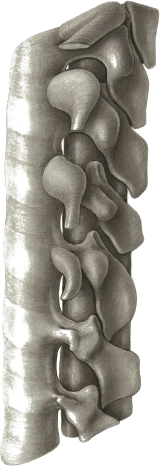
Link copiedSpinal involves narrowing of the spaces within your spinal canal, creating compression of neural structures. This narrowing can occur in the central canal (central stenosis) where the spinal cord or travels, or in the lateral recesses and foramina where individual exit.
The narrowing typically develops gradually through multiple mechanisms. changes in the discs can cause bulging into the spinal canal, while of the leads to formation and joint enlargement. The , which runs along the back of the spinal canal, can thicken and buckle inward, further reducing available space.
As these structures encroach on the neural space, they create a mismatch between the space available and the space needed for optimal neural function. The compression may be positional - worsening when your spine is extended and improving with flexion, which explains why many people find relief when leaning forward or sitting.
Overview
Contributing Factors
Link copiedYour spine's position significantly affects the amount of space available within the spinal canal. When you extend your back or stand upright, the canal diameter decreases due to buckling of the and narrowing of the lateral recesses. This positioning can worsen compression of already compromised neural structures.
Flexing your spine has the opposite effect - it increases canal dimensions by stretching the ligamentum flavum and opening up the lateral recesses. This is why many people with spinal naturally adopt a slightly flexed posture when walking or find relief when leaning on a shopping cart.
Walking on an incline often feels easier than walking on flat ground because the incline naturally puts you in a slightly flexed position. Similarly, cycling is usually tolerated better than walking because the cycling position maintains spinal flexion while allowing cardiovascular exercise.
Symptoms
Clinical Presentation
Link copiedPrimary Symptoms
Associated Symptoms
Typical pattern
The classic pattern I see is someone who can sit comfortably for long periods but develops leg symptoms within minutes of standing or walking. They often tell me they can walk much further in a grocery store while leaning on a cart, or that they can bike for miles but struggle to walk to the mailbox.
Symptoms
Differential Diagnosis
Link copiedConditions with similar presentations:
Vascular Claudication (Peripheral Arterial Disease)
Key differences: Leg cramping with walking that does not depend on spine position. Symptoms resolve with standing still, not with sitting or forward flexion. Reduced or absent distal pulses, skin changes, and a history of cardiovascular risk factors. Ankle-brachial index clarifies the diagnosis.
Lumbar Radiculopathy (Disc Herniation)
Key differences: Typically unilateral, leg pain worse with sitting, forward bending, coughing, or sneezing. Often younger patients than with . or slump test reproduce symptoms. Not typically eased by leaning forward on a cart.
Hip Osteoarthritis
Key differences: Groin and anterior thigh pain, reduced and painful hip internal rotation, and worsening with weight-bearing activities such as walking and standing. Pain is not clearly eased by forward flexion of the spine. Stair climbing is often particularly provocative.
Peripheral Neuropathy (e.g. Diabetic)
Key differences: Symmetrical, stocking-distribution numbness and tingling that is not position-dependent. Present at rest and often worse at night. Usually a context of diabetes or another systemic driver. Reflex and vibration changes on examination.
Trochanteric Bursitis / Gluteal Tendinopathy
Key differences: Lateral hip pain over the , worse lying on that side and with single-leg stance. Not typically described as and does not change with spine position.
Myelopathy (Cervical or Thoracic Cord Compression)
Key differences: Bilateral leg heaviness, coordination changes, positive Babinski or Hoffman signs, and bladder changes that are not clearly related to walking position. Requires prompt neurological work-up rather than -focused rehabilitation.
When to seek professional help
Research
Key Research & Evidence
Peer-reviewed studies supporting the treatment approach.
Finding
Supervised physiotherapy is associated with more patients reaching clinically meaningful improvement and a lower surgery rate at one year
Research details
A randomized controlled trial (Minetama et al., Clinical Rehabilitation 2021, 1-year follow-up) found that at 1 year more patients receiving supervised physiotherapy reached a clinically meaningful improvement on the Zurich Claudication Questionnaire than those doing a home exercise program alone, for both symptom severity (60.5% vs 32.6%) and physical function (55.8% vs 32.6%). The surgery rate at 1 year was also lower in the supervised group (7.0% vs 23.3%). The 6-week program was delivered twice weekly and combined supervised physiotherapy with a home exercise program
Clinical relevance
Supervised physiotherapy in the first 6 weeks can produce better short-term outcomes than unsupervised home exercise and is associated with a lower likelihood of surgery within a year, supporting a structured trial of supervised conservative care as a reasonable first step
Finding
Flexion-based and aerobic exercises, including cycling, feature commonly in exercise programs that help lumbar spinal stenosis
Research details
A 2023 systematic review and intervention component analysis (Comer et al., Clinical Rehabilitation) of 13 randomised trials reporting 23 exercise interventions delivered to 1,440 participants found that most interventions included supervision and flexion-based exercises. Components that featured more often in successful interventions included stretches, strengthening or trunk muscle exercises, aerobic fitness exercises (especially cycling), and psychologically informed approaches
Clinical relevance
Exercise programs for spinal stenosis should prioritize supervised flexion-based activities combined with cycling to maximize symptom relief and functional improvement, as flexion positioning opens the spinal canal and reduces neural compression
Finding
Manual therapy with exercise improves walking distance and reduces pain
Research details
Studies of combined manual therapy, exercise, and progressive body-weight-supported treadmill walking programs showed significant disability reduction and patient satisfaction after 6 weeks. Directed exercise and manual therapy proved superior to self-directed exercise for short-term walking capacity (mean difference 293.3 meters, 95% CI: 61.7-524.9), back pain (mean difference -1.1, 95% CI: -1.8 to -0.4), and leg pain (mean difference -0.9, 95% CI: -0.2 to -1.5)
Clinical relevance
Combining manual therapy with progressive weight-bearing exercise produces superior functional outcomes compared to exercise alone, with clinically significant improvements in walking tolerance and pain reduction supporting multimodal conservative management
Management
Evidence-Based Management
Treatment strategies with the strongest support in the current literature.
Primary approach
Flexion-biased exercise, strength work, and cardio (often cycling) meaningfully improve walking tolerance for most patients and can delay or avoid surgery
Complementary
Activity modification using positioning strategies and adaptive equipment allows maintained function while managing symptoms
Prevention & long-term
Early intervention with flexibility exercises and cardiovascular fitness can slow functional decline and maintain independence in daily activities
Detailed management strategies
Position Management
Learning to use spinal flexion positions during activities can significantly increase walking tolerance and reduce symptoms
Important precautions
- Avoid prolonged extension positions
- Use support when needed
Adaptive Equipment
Walking aids like canes or rollators can help maintain independence by providing support and encouraging beneficial postures
Important precautions
- Ensure proper fitting and training
- Don't delay use if it helps function
Activity Pacing
Breaking activities into manageable segments with rest periods allows completion of necessary tasks
Important precautions
- Plan activities around symptom patterns
- Don't push through severe symptoms
Alternative Exercise
Activities like swimming, cycling, or recumbent exercise can maintain fitness when walking is limited
Important precautions
- Choose activities that keep spine in neutral or flexed positions
- Monitor for any worsening symptoms
Management
Treatment Techniques
Evidence-based manual therapy and intervention approaches.
Treatment approaches supported by current research and clinical guidelines
Recommended treatment approaches
Treatment approaches are individualized to each patient's needs and goals. All interventions require explicit informed consent, and treatment plans are collaboratively modified based on your preferences and response to care.
Pain Education & Self-Management
Understanding pain science to reduce fear and improve movement confidence alongside active rehabilitation.
Joint Mobilization
Graded techniques to restore joint movement and reduce stiffness.
Post-Surgical Rehabilitation
Evidence-based recovery programs following surgery to restore function and strength.
Rehabilitation
A Typical Rehabilitation Progression
Three phases, from settling symptoms to returning to full activity.
Recovery from Spinal Stenosis is usually staged: calm the symptoms first, then rebuild the strength and capacity of the area, then return to your full activities. The three phases below show the kind of progression the evidence supports and that I commonly work through in clinic. They are here to show you what the road can look like, not to act as a personal program.
- Phase 1
Foundation: Open the Canal, Rebuild Walking Tolerance
The priority early on is teaching the spine position that reduces symptoms (slight flexion), restoring confidence with walking in small doses, and starting flexion-biased cardio. Guideline synthesis from Bussières et al. (J Pain 2021) and the NASS CPG supports supervised exercise, , and education as first-line care.
Examples, not a prescription
- Interval walking: walk until symptoms are mild (2 to 3/10), sit or lean on a counter for 1 to 2 minutes, repeat for 10 to 15 minutes total
- Stationary cycling in a slightly flexed position, starting with 10 to 15 minutes at low resistance
- Single and double knee-to-chest in supine, 30 seconds, 2 to 3 repetitions, for segmental flexion
- Pelvic tilts and cat-camel on hands and knees, 10 slow repetitions
- Education on leaning on a cart, using a walker if needed, and breaking up standing tasks
Ready to progress when
Walking intervals up to 10 minutes before needing to sit, comfortable 20-minute cycle, and confident use of flexion-based position strategies during daily tasks.
- Phase 2
Progressive Loading: Build Hip, Trunk, and Cardiovascular Capacity
Once baseline tolerance improves, the focus shifts to strengthening the hips and trunk, which allows the spine to stay in a more tolerant position during longer walks, and extending cardiovascular capacity so fitness is not the limiting factor.
Examples, not a prescription
- Glute bridges and clamshells, 2 to 3 sets of 10 to 12
- Sit-to-stand repetitions from a sturdy chair, 2 to 3 sets of 8 to 10
- Step-ups onto a low step, 2 to 3 sets of 8 per side
- Dead bug and modified bird dog for trunk control, 2 to 3 sets of 6 to 8
- Progressive cycling to 20 to 30 minutes, or aquatic walking if available
Ready to progress when
20 to 30 minute walking tolerance with planned breaks, tolerating basic resistance work without leg symptom flare, and able to manage a full shopping trip or equivalent errand.
- Phase 3
Return to Function: Maintain Independence and Capacity
The final phase is about sustainability: maintaining enough hip strength, trunk capacity, and cardiovascular fitness to keep independence in daily activities and reduce the risk of functional decline. often progresses slowly, so the maintenance plan matters as much as the rehab plan.
Examples, not a prescription
- Longer continuous or interval walks, built up gradually to 30 to 45 minutes with planned flex breaks
- Progressive lower body resistance work (goblet squat, leg press, hip hinge with support) at moderate loads
- Ongoing cycling two to three times weekly at moderate intensity
- Balance and single-leg stance work to reduce fall risk, 2 to 3 sets of 20 to 30 seconds per side
- A written maintenance programme the patient can follow independently, reviewed periodically
Ready to progress when
Full independence with daily activities, walking tolerance that matches the patient's functional goals, and a sustainable home programme the patient is willing and able to continue.
Management
Prognosis & Recovery
What outcomes and recovery factors typically look like.
Expected timeline
Symptoms often remain stable or progress slowly over years; significant improvement with conservative care is possible in 6-12 weeks
Natural history
often progresses slowly; some people maintain stable function for years while others experience gradual decline requiring surgical intervention
Factors affecting recovery
Management
Measuring Progress
How to track the recovery arc week to week.
Day-to-day tracking
I track your walking distance before symptoms begin, how long you can stand before needing to sit, and your confidence with daily activities that require mobility
Assessment tools
Swiss Spinal Stenosis Questionnaire to monitor symptom severity and functional impact over time
Activity targets
Maximizing your walking capacity and maintaining independence with daily activities
Management
Frequently Asked Questions
Common concerns and answers about this condition.
Does spinal stenosis always require surgery?
Does spinal stenosis always require surgery?
No. Many people manage well without surgery. The NASS clinical guideline on spinal stenosis and the 2021 multi-disciplinary CPG (Bussières et al., J Pain 2021) both recommend a structured trial of conservative care as the first step. Surgery is considered when leg symptoms are severely limiting walking and quality of life despite appropriate non-surgical management, or when there is progressive neurological loss.
Why can I bike for an hour but barely walk a block?
Why can I bike for an hour but barely walk a block?
Because of spine position. Cycling keeps the slightly flexed, which opens the spinal canal and unloads the nerves. Walking upright closes it down again, which is why symptoms come on within minutes of standing or walking and ease when you sit or lean on a cart. This position-dependent pattern is characteristic of .
How far will my walking distance improve with physiotherapy?
How far will my walking distance improve with physiotherapy?
Modest but meaningful gains are realistic for most people. A supervised flexion-biased exercise and programme, combined with cycling or similar flexed-position cardio, can meaningfully improve walking tolerance within 6 to 12 weeks. The programme does not reverse the narrowing itself. It improves how well you function inside it by improving strength, endurance, and tolerance.
Should I avoid walking because it brings on symptoms?
Should I avoid walking because it brings on symptoms?
No. Interval walking works better than avoidance. Walk until symptoms are mild, sit or lean forward until they ease, then go again. Over weeks, the distance before symptoms usually extends. Avoidance leads to deconditioning, which makes everything harder.
Is leaning on a shopping cart a sign that I am getting worse?
Is leaning on a shopping cart a sign that I am getting worse?
No. It is a sensible adaptation. Leaning forward opens the spinal canal and lets you walk further. Using a walker or a rollator with handles does the same thing, and using one is not a step backward. Maintaining activity matters more than how upright you look doing it.
Will an epidural steroid injection help?
Will an epidural steroid injection help?
Sometimes, and usually short-term. Injections can reduce leg symptoms enough to participate in rehabilitation, but the effect is typically measured in weeks to a few months rather than years. They are reasonable to consider when pain is limiting your ability to progress with exercise, but they are not a standalone solution.
What symptoms mean I should see a doctor urgently?
What symptoms mean I should see a doctor urgently?
New or progressive leg weakness, foot drop, loss of bladder or bowel control, saddle-area numbness, or a sudden change in balance. These can indicate or significant neurological compromise and need urgent medical assessment rather than physiotherapy as a first step.
Related Conditions
Conditions I commonly see alongside, or confused with, this one.