Meniscal Injuries
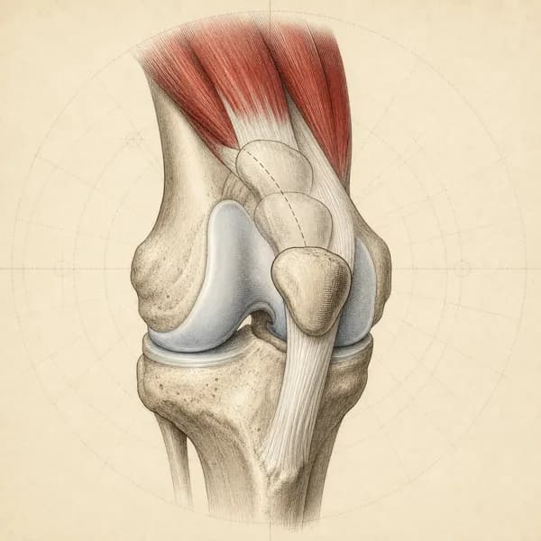
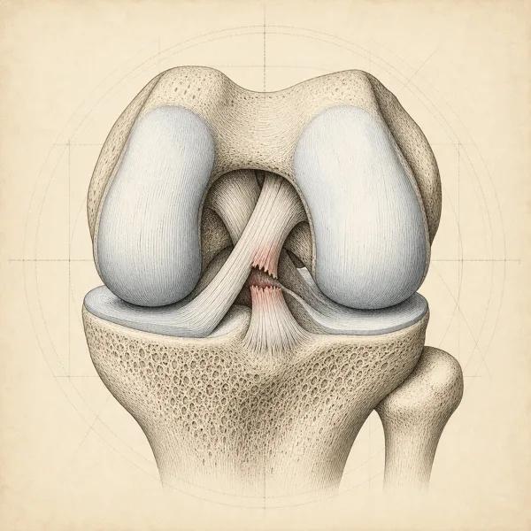
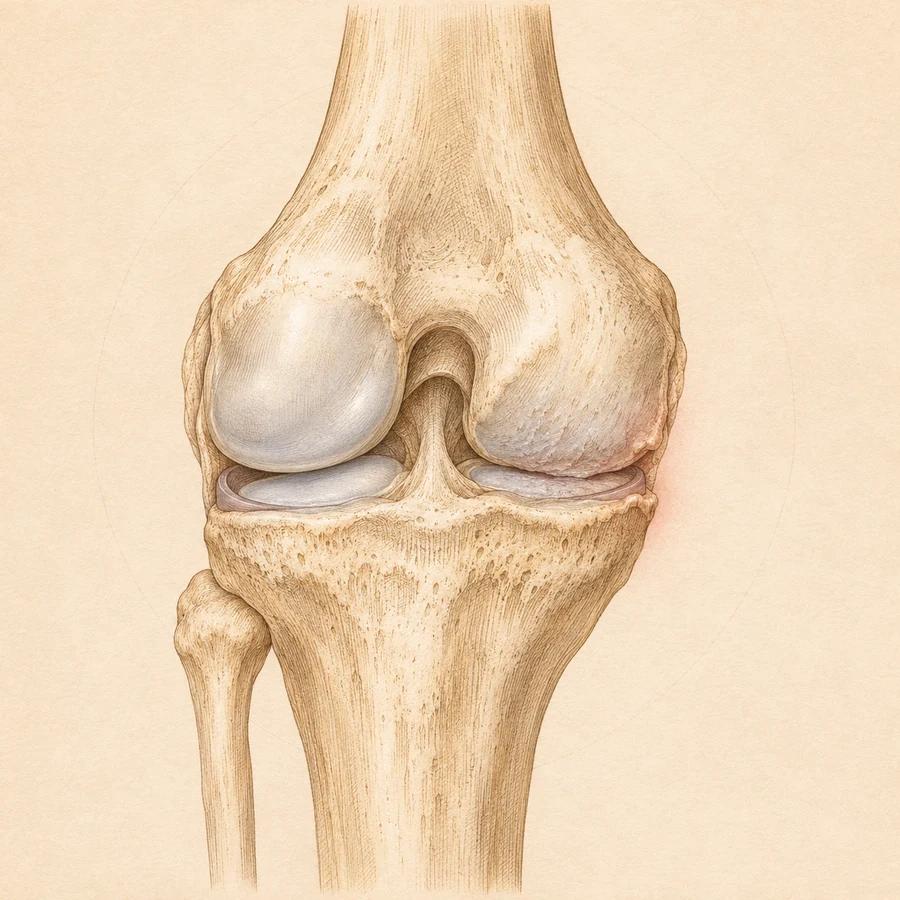
Each knee has two menisci: cartilage pads that help distribute load between the thigh bone and shinbone. Tears can follow a twist or develop gradually with age. They may cause pain, swelling or catching, but some cause no symptoms. A knee that truly locks needs medical assessment before rehabilitation.
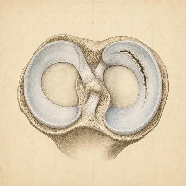
Does this sound like you?
- Pain along the joint line (the gap between the thigh bone and shin bone)
- Clicking or catching sensation
- Swelling after activity
- Difficulty fully straightening the knee
- Pain with twisting movements
Often alongside it
- A feeling of the knee giving way
- Stiffness after sitting
- Difficulty squatting
- Night pain
- Weakness in the front thigh muscle (quadriceps)
Tears from an injury (traumatic tears) start suddenly with a twist. Tears from gradual wear (degenerative tears) develop slowly. Catching and similar mechanical symptoms are common.
Before using this comparison, check when these symptoms need a doctor first.
When to see a doctor first
A hot, red or very swollen knee with fever, chills or feeling generally unwell
Go to emergency now. This can be a joint infection.
After surgery: any of these and you cannot reach your surgical team, or you feel very unwell
Go to emergency
Sudden shortness of breath or chest pain, coughing up blood or feeling faint, especially with a painful or swollen calf
Go to emergency now, or call 911. A clot that has travelled to the lungs (pulmonary embolism) needs to be ruled out
The knee is locked and will not straighten, especially after a twist
Possible large tear that has flipped into the joint (a displaced, bucket-handle tear). Get it assessed the same day by your family doctor, urgent care or emergency; it needs an urgent orthopaedic referral
Large swelling within a few hours of an injury, or you cannot put weight on the leg
Possible ligament injury or fracture. Same-day medical assessment
A newly hot, red or very swollen knee without fever, chills or feeling generally unwell
Get medical assessment today to rule out a joint infection. Infection is possible even without a fever.
Fever with knee pain but no new heat, redness or swelling in the knee
Get medical assessment today to rule out a joint infection.
After surgery: calf pain or swelling, a wound that is red, hot or leaking, or a fever
Possible blood clot or infection. Contact your surgical team the same day
How it is treated
For tears from gradual wear in middle-aged and older knees (degenerative tears), trials found knee function after physical therapy was comparable to keyhole surgery (METEOR, ESCAPE), and surgery did no better than a placebo operation (FIDELITY). For these tears, structured exercise is the usual first step. For a tear from an injury in a younger, active person, many tears that do not block the knee's movement also respond well to exercise; a knee that locks and will not straighten needs medical assessment the same day, and whether surgery is needed is decided case by case with an orthopaedic surgeon.
Balance and control training (neuromuscular training) and strengthening the front thigh muscle (quadriceps) improve knee control and can reduce catching and giving way.
Keeping up quadriceps and hip strength after rehab helps the knee cope with load and can keep symptoms from returning.
A typical rehabilitation progression
Recovery from meniscal injuries is usually staged: calm the symptoms first, then rebuild the strength and capacity of the area, then return to your full activities. The phases below show how that can progress.
Phase 1 · Weeks 1 to 3
Settle Symptoms and Protect Range
Bring swelling and pain to a manageable level, get the knee fully straight again, and start switching the front thigh muscle (quadriceps) back on without irritating the joint line. Getting the knee fully straight early is important, since a knee that still will not straighten can lead to an awkward walking pattern and a thigh muscle that stays switched off for longer.
Example exercises
- Quadriceps setting (tightening the thigh muscle with the leg straight) and straight-leg raise, 3 sets of 10 to 12, focusing on getting the knee fully straight
- Heel slides and bending the knee while lying on your back, as far as is comfortable, 2 to 3 sets of 10
- Short-arc quadriceps extension (straightening the knee from a slight bend, with a roll under the knee) through a pain-free range close to straight, 3 sets of 10 to 12
- Side-lying (lifting the top leg while lying on your side) and clamshell, 3 sets of 12 to 15 per side
- Stationary cycling with high saddle and light resistance, 10 to 15 minutes
Phase 2 · Weeks 3 to 8
Building Load and Single-Leg Strength
Rebuild real lower-body strength with exercises where your foot stays on the ground (closed-chain), add single-leg work, and bring back controlled twisting. The ESCAPE trial supports exercise-based physiotherapy for tears, and METEOR found no clear intention-to-treat advantage from surgery plus physiotherapy over physiotherapy alone in adults aged 45 or older with a tear and mild to moderate .
Example exercises
- Goblet squat (holding a weight at your chest) to a box, 3 to 4 sets of 8 to 10, gradually lowering box height and adding load
- Leg press, 3 sets of 8 to 12, progressively loaded
- Step-ups and controlled step-downs, 3 sets of 8 to 10 per side
- Single-leg Romanian deadlift (hinging forward at the hip on one leg), 3 sets of 8 per side, for back-of-thigh (hamstring) strength and control of the pelvis
- Spanish squat (a squat with a strap behind the knees) or a heavy, slow leg extension to strengthen the front of the thigh, 3 sets of 8 to 10
Phase 3 · Weeks 8 to 16+
Return to Sport, Cutting, and Higher Loads
Rebuild the ability to absorb and produce force when twisting and moving fast. This phase is essential for people returning to running, team sports or recreational activities that load the knee, and is where many unfinished rehab programs fall short.
Example exercises
- Hops on two legs, then on one leg, 3 to 4 sets of 5 to 8
- Jumping and landing drills () such as drop jumps and bounds, with attention to how you land
- Change-of-direction drills at graded speeds, building from low to moderate to high intensity
- Heavier squats and deadlifts on both legs, matched to your training goals
- Graded return-to-run or sport-specific program, increasing volume gradually and adjusting each step to how the knee responds over the following 24 hours
What you can do now
- Ease off twisting and deep loaded squatsSharp twisting, deep squats with weight early on and sudden big jumps in training tend to flare symptoms. Cutting back on these for a few weeks, while you rebuild strength, lets the knee settle.This advice is for a tear assessed as stable or from gradual wear. Root tears, displaced tears, a new major injury and a repaired meniscus need different advice.
- Keep strengthening your thigh and hipLoading the front thigh (quadriceps) and hip muscles is part of the treatment. Many tears from gradual wear stop causing symptoms with strengthening, load management and time, even if the tear stays visible on a scan.
- Add load back in graduallyOnce symptoms settle, add activity back in step by step. With a tear assessed as stable or from gradual wear, running or training is often possible with these changes.
- Tell catching apart from true lockingClicking, catching or a knee that hesitates is common with tears from gradual wear and is usually managed without surgery. True locking is different: the knee physically will not straighten fully and you cannot unlock it yourself.If the knee is locked and will not straighten, especially after a twist, get it assessed the same day by your family doctor, urgent care or emergency for an urgent orthopaedic referral. Large swelling within a few hours of an injury, or not being able to put weight on the leg, needs a same-day medical assessment.
What I use in clinic
- Therapeutic ExerciseStrong muscles help protect the knee and reduce symptoms, so the load is built up step by step. For tears from gradual wear (degenerative tears), knee function after physical therapy was comparable to keyhole (arthroscopic) surgery in randomised trials.
- Balance and Control Training (Neuromuscular Training)Balance and control training improves knee stability and reduces symptoms.
Treatments on this site
Book an assessment for meniscal injuriesNextRecovery timeHow long it takes
- Weeks 1 to 3Settle Symptoms and Protect Range
- Weeks 3 to 8Building Load and Single-Leg Strength
- Weeks 8 to 16+Return to Sport, Cutting, and Higher Loads
Most people improve within 6 to 12 weeks with appropriate rehabilitation.
Many tears stop causing symptoms with time and appropriate management.
What changes the timeline
- The type of tear and where it is
- Age and activity level
- Arthritis in the same knee
- Keeping up with the exercises
Progress. I track how much the pain gets in the way of the tasks that matter to you, how well you move when I test those tasks in clinic, and how confident you feel in daily activities
Conditions that can feel similar
- Knee Osteoarthritis
- Pain that comes on gradually when you stand and walk on the leg, morning stiffness under 30 minutes, grinding or crackling in the joint (crepitus), and age typically over 45. It frequently occurs alongside wear-related meniscus tears, and the two are often best managed as one rehab problem rather than separately.
- ACL or MCL Injury
- A clear injury, often with an audible pop, swelling within hours, and a sense of the knee being unstable or giving way when changing direction or twisting. Tests of how far the shin bone moves forward (the Lachman and anterior drawer tests) help identify an ACL injury, and those findings are not typical of a meniscus tear on its own.
- Patellofemoral Pain
- Pain behind or around the kneecap rather than along the joint line, aggravated by stairs and long sitting with bent knees, without true catching or a twisting injury.
- Pes Anserine Bursitis or Tendinopathy
- Tenderness in one spot on the inner shin bone, 5 to 7 cm below the joint line, not at the joint line itself. It is often mistaken for an inner meniscus problem, particularly in middle-aged people.
- Medial Plica Syndrome
- A tender band that can be felt over the inner end of the thigh bone (the medial femoral condyle), with clicking or snapping as the knee bends and straightens. It often looks like an inner meniscus problem, but twisting with weight on the leg does not bring it on.
- Referred Pain from the Hip
- Pain at the front or inner side of the knee, with turning the hip inward limited or painful, and a hip test that brings on pain (FADIR). A hip problem showing up as knee pain is commonly missed, especially in older adults.
Side by side
NextQuestionsWhat patients ask me most
Usually not. The FIDELITY trial (Sihvonen et al., NEJM 2013) randomly assigned 146 patients with wear-related (degenerative) tears of the inner meniscus and no osteoarthritis to keyhole surgery that trims the tear (arthroscopic partial meniscectomy) or to placebo (sham) surgery, and found no difference in any primary outcome at 12 months. The ESCAPE trial (van de Graaf et al., JAMA 2018) compared physical therapy with arthroscopic partial meniscectomy in 321 patients with degenerative tears. Its 5-year follow-up (Noorduyn et al., JAMA Network Open 2022) found physical therapy was still no worse than surgery (noninferior) for knee function as patients rated it. For degenerative tears, a structured rehabilitation program is a reasonable first step for many people.
Traumatic tears usually happen in a specific moment, often a loaded twist or cutting movement in a younger athletic knee, with immediate pain, swelling, and sometimes a true locking episode. Degenerative tears develop over years in middle-aged and older knees, often without a clear injury event, and commonly sit alongside early osteoarthritis. The rehabilitation pathway is quite different, which is why the history matters as much as the MRI.
The outer third has a blood supply and some capacity to heal, particularly in younger people with tears near the outer edge (peripheral tears). Healing potential varies with the tear's location, pattern and treatment. The inner region has much less blood supply, so it has less ability to heal on its own. Symptoms can still settle: plenty of degenerative tears stop causing symptoms with strengthening, load management and time, even if the tear itself stays visible on imaging.
Possibly, but true locking and a vague sense of catching are different. True locking means something in the joint physically stops the knee straightening fully and you cannot unlock it yourself, often from a large tear that flips into the joint (a bucket-handle tear). A knee that is locked and will not straighten needs assessment the same day by your family doctor, urgent care or emergency, and an urgent orthopaedic referral. A feeling of clicking, catching, or the knee 'hesitating' is common in degenerative tears and osteoarthritis, and is usually managed without surgery.
The METEOR trial (Katz et al., NEJM 2013) randomly assigned 351 patients with a meniscus tear and osteoarthritis to keyhole surgery to trim the tear (arthroscopic partial meniscectomy) plus physical therapy, or to physical therapy alone, and found no meaningful difference in function at 6 months. Thorlund and colleagues' 2015 BMJ systematic review and meta-analysis of keyhole surgery for knees with wear-related (degenerative) problems concluded that any benefit over non-surgical care was small, short-lived, and gone by one to two years. Physiotherapy is a reasonable first choice for most degenerative tears, and plenty of injury-related tears that do not block the knee's movement also respond well to loading.
A knee that is truly locked and will not straighten needs same-day medical assessment and an urgent orthopaedic referral. A displaced bucket-handle or flap tear can need surgical assessment. Root tears also deserve their own discussion because the attachment helps the meniscus distribute load. Repair, trimming and non-surgical care are different options; cartilage changes, alignment, tissue quality and the demands of recovery affect suitability. Tears near the outer edge with a useful blood supply may also be considered for repair. These choices are made with an orthopaedic surgeon.
Often yes, with modification, for a tear that has been assessed as stable or wear-related. Appropriately adjusted activity is often possible, and loading the front thigh and hip muscles is part of the treatment. Root tears, displaced tears, a new major injury and a repaired meniscus need different advice. Sharp twisting, deep squats with weight early on, and sudden large jumps in volume tend to flare symptoms. The usual approach is reducing aggravating activities for a few weeks while rebuilding strength, then adding load back in gradually.
Meniscus loss is a known risk factor for later knee osteoarthritis, and trimming the meniscus (partial meniscectomy) removes tissue that will not grow back. This is one of the reasons the evidence has pushed back against routine surgery for degenerative tears. Operating on a tear that would have settled with rehab can be counterproductive over decades. Keeping as much working meniscus as possible is generally the priority.
A root tear involves an attachment to the shin bone, while extrusion describes the meniscus extending outward beyond the bone’s edge. The assessment brings those findings together with symptoms, cartilage and leg alignment. A root repair has a different protection plan from a meniscus trim or rehabilitation for an assessed stable tear (Monson and LaPrade, 2025).
The science of meniscal injuries
The is that cushions and stabilizes the knee. Tears can be traumatic (usually in younger people) or (older adults). Degenerative tears are often part of early .
The outer third has blood supply and can heal, while the inner portion has poor healing capacity.
tears happen through two distinct mechanical patterns. Traumatic tears in younger athletes typically occur during cutting or pivoting movements when the foot is planted and the knee twists, trapping the meniscus between the thigh bone and shin bone. The risk is higher when the knee is slightly bent and rotates under load, as when you plant your foot to change direction in sports.
tears follow a different pattern and are much more common after age 40. These develop gradually from repetitive compressive and rotational forces during normal activities. With age, the meniscus becomes less elastic and more prone to tearing from everyday movements like squatting, kneeling, or even getting up from a low chair. Previous knee injury and repeated occupational kneeling or squatting are associated with meniscal tears.
I assess movement, hip and thigh strength, knee mobility and your activity demands to choose a manageable rehabilitation plan. Some people have hip weakness or an inward movement of the knee during loaded tasks, and those findings may guide exercise choices. I also address restricted movement when it limits a useful activity. Once the meniscus is damaged, it can catch or pinch during normal knee movement, creating mechanical symptoms like clicking, locking, or catching.
What the studies show
- 2022
Physical therapy compared with keyhole surgery for tears from gradual wear: 5-year follow-up of the ESCAPE trial
In 321 people aged 45 to 70 with a meniscus tear from gradual wear (a degenerative tear), physical therapy was still not worse than keyhole surgery to trim the tear (arthroscopic partial meniscectomy) for knee function, as people rated it themselves, at 5 years. Knee osteoarthritis progressed at a similar rate after both treatments.
Supports a structured physical therapy program as the first step for most tears from gradual wear in people of this age.
Noorduyn JCA, van de Graaf VA, Willigenburg NW, et al. Effect of physical therapy vs arthroscopic partial meniscectomy in people with degenerative meniscal tears: five-year follow-up of the ESCAPE randomized clinical trial. JAMA Netw Open. 2022;5(7):e2220394. PMID: 35802374.
Keep reading
Guides on knee pain
Related conditions
 ACL InjuriesOften occur together; both involve knee stability and similar injury mechanisms
 MCL/LCL SprainsMeniscus tears can occur with ligament injuries in complex knee trauma
 IT Band SyndromeBoth can cause lateral knee pain and may be confused diagnostically
 Patellar Tendinopathy (Jumper's Knee)Meniscus and patellar tendon problems can both cause knee pain, but location, symptoms and examination help distinguish them
I write and review every page on this site myself, and the review date at the top is the last time I checked this one. If something here is out of date, tell me.
Getting back to it
Physiotherapy for meniscal injuries, built around the activity you want back.