
Plantar Calcaneal Spur (Heel Spur)
A plantar calcaneal spur is a small ledge of extra bone on the underside of the heel bone, seen on a side view X-ray. Heel spurs often show up in people who have no heel pain. The phrase describes the shape of the bone; on its own it does not explain the pain or decide the treatment. This guide covers what the wording on the report means, what it does and does not tell you, and what the treatment guidelines recommend.
Assessing and treating foot and ankle pain at the Burlington clinic. Convenient for Waterdown, Oakville, Hamilton, Flamborough, and Carlisle residents.
When to see a doctor first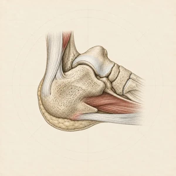
What “plantar calcaneal spur” means on an X-ray report
What it is. The calcaneus is the heel bone. A plantar calcaneal spur is a small outgrowth of bone on its underside, pointing forward toward the arch. It shows on a side view of the foot or ankle, usually taken standing. It usually grows roughly parallel to the ground rather than down into the sole.
Where it sits. A spur is often pictured growing where the plantar fascia pulls on the heel. In anatomy studies, most spurs sat just above the plantar fascia, among the attachments of the small muscles of the sole, and only a few sat inside the fascia itself (Abreu et al., 2003; Kumai and Benjamin, 2002). The internal structure of the bone lines up with the load of standing and walking rather than with the pull of the fascia, which has led researchers to read the spur as a response to load through the heel (Li and Muehleman, 2007; Menz et al., 2008). The samples were small, and the cause is still debated.
The words that come with it. Reports name the same finding several ways and add a few standard words. These are the ones you are most likely to see.
| On the report | What it means |
|---|---|
| Plantar calcaneal spur, plantar heel spur, inferior or subcalcaneal spur | Bony outgrowth on the underside of the heel bone. All of these name the same finding. |
| Calcaneal enthesophyte or plantar calcaneal enthesophyte | The same finding, named after where it grows. An enthesis is the place where a tendon, ligament or fascia attaches to bone. |
| Calcaneal spurring | The same finding. “Spurring” is the usual radiology wording. |
| Is noted, is seen, is present, is evident | The radiologist saw it. On their own, these words do not signal concern or urgency. |
| Small, moderate or large | The reader’s own description of size. There is no standard cut-off. Research sometimes sorts spurs as under 5 mm, 5 to 10 mm and over 10 mm (Ahmad et al., 2016). |
| A measurement in millimetres | The length of the spur from its base to its tip on the side view. |
| Bilateral | Spurs on both heels. Common, especially with age. |
| Degenerative or enthesopathic change | Change at the attachment that tends to come with load and age. It is not a diagnosis on its own. |
| Posterior, dorsal or Achilles enthesophyte | A spur at the back of the heel, where the Achilles tendon attaches. A different place and a different problem, covered in the next section. |
| Plantar fascia thickening (ultrasound or MRI) | The fascia is thicker than usual where it attaches to the heel. More than 4 mm is the usual research threshold (McMillan et al., 2009). It often comes with a spur. |
Plantar calcaneal spur, plantar heel spur, inferior or subcalcaneal spur
Bony outgrowth on the underside of the heel bone. All of these name the same finding.
Calcaneal enthesophyte or plantar calcaneal enthesophyte
The same finding, named after where it grows. An enthesis is the place where a tendon, ligament or fascia attaches to bone.
Calcaneal spurring
The same finding. “Spurring” is the usual radiology wording.
Is noted, is seen, is present, is evident
The radiologist saw it. On their own, these words do not signal concern or urgency.
Small, moderate or large
The reader’s own description of size. There is no standard cut-off. Research sometimes sorts spurs as under 5 mm, 5 to 10 mm and over 10 mm (Ahmad et al., 2016).
A measurement in millimetres
The length of the spur from its base to its tip on the side view.
Bilateral
Spurs on both heels. Common, especially with age.
Degenerative or enthesopathic change
Change at the attachment that tends to come with load and age. It is not a diagnosis on its own.
Posterior, dorsal or Achilles enthesophyte
A spur at the back of the heel, where the Achilles tendon attaches. A different place and a different problem, covered in the next section.
Plantar fascia thickening (ultrasound or MRI)
The fascia is thicker than usual where it attaches to the heel. More than 4 mm is the usual research threshold (McMillan et al., 2009). It often comes with a spur.
When to see a doctor first
Get seen today
Heel pain after a fall, a jump from a height or a snap or pop, or you cannot walk or stand on tiptoe
Seek same-day medical assessment and keep weight off the foot until it is checked.
A newly hot, red or swollen heel or foot, with or without a fever, or feeling unwell
Seek same-day medical assessment to rule out infection, or go to emergency if you feel unwell.
Diabetes and a new wound, blister or change in skin colour on the foot
Get it seen today by your doctor or diabetes foot care team. Foot problems can be more serious with diabetes.
Check with your doctor
Pain that keeps building after more walking or running, or a move to harder ground, with pain when the sides of the heel are squeezed
See your doctor to rule out a stress fracture of the heel bone before loading it further.
Burning, tingling or numbness in the heel or sole
See your family doctor. A trapped nerve in the foot, or a nerve from the lower back, needs to be checked.
Pain in both heels with morning stiffness over half an hour, back pain at night, or other swollen joints
See your family doctor to check for an inflammatory arthritis.
A history of cancer, unexplained weight loss, or constant pain at night that is not linked to activity
See your family doctor for a medical workup before starting physiotherapy.
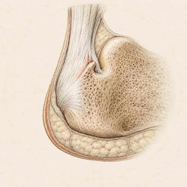
A spur at the back of the heel is a separate finding
Where it sits. A posterior, dorsal or Achilles enthesophyte grows where the Achilles tendon attaches to the back of the heel bone, pointing up into the lowest fibres of the tendon. It is a different place from a spur under the heel, and a different problem.
What it goes with. It is also common in people without symptoms. When the back of the heel hurts, it goes with insertional Achilles tendinopathy, which has its own plan. The Achilles tendinopathy page covers it. Reports often mention spurs at both sites together.
What it does and does not tell you
It is common without pain. How often spurs turn up depends on age and on who was X-rayed. In one study of people with pain in one heel, spurs were found in 85 percent of the painful feet and in 72 percent of the painless feet on the other side (Okcu et al., 2023). Spurs become more common with age and with higher body weight (Menz et al., 2008).
It travels with heel pain, but is not shown to cause it. People with heel pain on the underside of the heel are more likely to have a spur. A review of the imaging studies judged this “an association only and unlikely to be the cause of pain”, and found the spur of limited relevance to treatment unless it is fractured (Drake et al., 2022).
It rarely comes alone. In adults over 50 with foot pain, a spur on its own was found in 6 percent of feet. Much more often it sat alongside a thickened plantar fascia (Menz et al., 2019).
Size is not a reliable guide on its own. Some studies link longer spurs with more pain and others find no link, and in some treatment studies larger spurs improved more. Size has not shown a consistent link with how much it hurts or how well it recovers.
It did not change the long-term outlook. In a study that followed people with plantar fasciitis for 5 to 15 years, having a spur at the start made no difference to how they recovered (Hansen et al., 2018).
It usually stays on the X-ray. In treatment studies the spur mostly looked the same afterwards while the pain improved (Lee et al., 2003; Yalcin et al., 2012). Heel pain can improve without the spur changing.
What the treatment guidelines recommend
Treatment is aimed at the painful heel, most often plantar fasciitis, not at the spur. The guidelines below do not change the plan because a spur is present (Koc et al., 2023; Morrissey et al., 2021).
- Education and load. Understanding what the spur is and is not, and adjusting how much standing, walking and running the heel takes for a while, are part of core care in a best practice guide built from trials, clinicians and patients (Morrissey et al., 2021).
- Stretching and taping. Plantar fascia and calf stretching and taping of the foot carry the strongest grade in the physical therapy guideline for heel pain, taping for short-term relief alongside other care (Koc et al., 2023).
- Strengthening. The guideline recommends resistance training for the foot and ankle muscles. In one trial, high-load heel raises improved function faster than stretching at three months, with no difference at one year (Rathleff et al., 2015).
- Hands-on care. Joint mobilization and soft tissue work for the foot, ankle and calf are recommended alongside exercise to ease pain and restore movement (Koc et al., 2023).
- Night splints and insoles. Night splints are recommended for people who keep getting pain with the first steps in the morning. Insoles and heel cups are not recommended on their own for short-term relief, but can be added to other care (Koc et al., 2023).
- Shockwave. Shockwave can be effective for heel pain that has not improved with core care, on low to moderate certainty evidence (Morrissey et al., 2021). Results were similar with and without a spur in several studies, and it does not reliably remove the spur.
- Injections. A steroid injection may slightly reduce heel pain for about a month, on low quality evidence, and rare risks include rupture of the plantar fascia (David et al., 2017). It is arranged through your doctor.
- Surgery. Considered for a small number of people after a long period of well-run non-surgical care, and decided with a surgeon. Removing the spur is not usually the aim, and in one series adding spur removal to fascia surgery was linked with slower recovery (Cooperman et al., 2025).
When the report deserves a closer look
- The report describes the spur as fractured or broken. That is rare, and it is managed as a fracture.
- It describes an ill-defined spur, erosions, or spurs at several attachments, or mentions an inflammatory arthritis. That pattern can point to a reactive or psoriatic arthritis and needs a doctor’s assessment.
- An MRI shows bone marrow oedema through the body of the heel bone, which can be a stress reaction, or a tear of the plantar fascia.
- An MRI mentions wasting of a small muscle on the outer side of the sole (abductor digiti minimi), which can go with irritation of a nerve under the heel (Chundru et al., 2008).
- The report flags something unrelated to the spur elsewhere in the bone. Ask the doctor who ordered the X-ray to go through it with you.
- The pain does not follow the usual pattern of first-step pain under the heel, or has not improved after a fair trial of well-run care. Imaging and other diagnoses are worth reconsidering then (Koc et al., 2023).
How I approach a heel spur in clinic
I begin with the heel itself: where exactly it hurts, whether the first steps in the morning or after sitting are the worst, how long you are on your feet in a day and on what surfaces, what shoes you wear, what changed before it started, and what you have already tried. I also ask what you were told about the spur, because the picture of a sharp point of bone can make people wary of walking on or loading the foot.
On examination I check where the tenderness is, test ankle and big toe movement, calf strength with single-leg heel raises, balance and how you walk. I screen for a stress fracture, a trapped nerve and a problem at the back of the heel, and for the lower back when the symptoms suggest it. I record a few measures I can repeat so you can see whether the plan is working.
The plan usually centres on the painful heel and can include explaining what the spur means, adjusting load for a while, calf and plantar fascia stretching, taping for short-term relief, and progressive strengthening of the calf and foot. Joint mobilization, soft tissue work and dry needling to the calf can sit alongside the exercise when they help. If the heel has not improved with that, shockwave can be a reasonable next step, and I can coordinate with your family doctor when an injection or a surgical opinion is worth discussing.
Heel spur questions I hear most
What does a plantar calcaneal spur mean on an X-ray?
A small outgrowth of bone on the underside of the heel bone, seen on the side view. It is common in people without heel pain, and it describes the shape of the bone rather than the cause of any pain.
What does “small plantar calcaneal spur is noted” mean?
“Noted” means the radiologist saw it, and “small” is their own description of size. There is no standard cut-off. A small spur is a common finding and does not by itself mean anything needs to be done.
Is a heel spur the same as plantar fasciitis?
No. They often turn up together, but a heel spur is a finding on the bone and plantar fasciitis is irritation of the thick band under the foot. People with heel pain are more likely to have a spur, but the spur is not shown to cause the pain.
Do heel spurs cause pain?
The evidence does not show that a spur causes pain on its own. Spurs are common in painless feet, and having one did not change long-term recovery in a 5 to 15 year study. The pain usually comes from the tissues around the heel.
Can a heel spur go away?
The spur usually stays on the X-ray. In treatment studies it mostly looked the same afterwards while the pain improved, so the aim is a heel that hurts less and works better, rather than a different X-ray.
What is the difference between a calcaneal enthesophyte and a heel spur?
They are two names for the same thing. “Enthesophyte” is the radiology word for bone growing where a tendon, ligament or fascia attaches. “Heel spur” is the everyday name for the same thing. An osteophyte is different: it forms at the edge of a joint.
What is a posterior calcaneal enthesophyte?
A spur at the back of the heel, where the Achilles tendon attaches. It is common without symptoms, and when it hurts it goes with insertional Achilles tendinopathy, which is managed differently from heel pain under the foot.
Does stretching help a heel spur?
Calf and plantar fascia stretching are recommended for heel pain under the foot. They treat the painful tissue, not the spur, which usually stays the same.
Does shockwave therapy work for heel spurs?
Shockwave can be effective for heel pain that has not improved with core care. It does not reliably remove the spur, and results were similar with and without a spur in several studies.
Do I need surgery to remove a heel spur?
Usually not. Removing the spur is not usually the aim of heel pain surgery, which is considered only after a long period of well-run non-surgical care, and decided with a surgeon.
Do I need an MRI or ultrasound?
Usually not. Imaging is generally not needed when the symptoms fit plantar fasciitis, unless care is not working or something else is suspected, such as a stress fracture, a tear or a trapped nerve.
Evidence this page is built on
The main sources behind this guide. Other studies are named in the text where they are used.
Heel Pain - Plantar Fasciitis: Revision 2023
Clinical practice guideline. Recommends manual therapy, plantar fascia and calf stretching and taping (strongest grade), resistance training for the foot and ankle, night splints for first-step pain, and orthoses only alongside other care. Imaging is usually not needed for typical plantar fasciitis unless care is not working.
Management of plantar heel pain: a best practice guide informed by a systematic review, expert clinical reasoning and patient values
Core care is taping, stretching and individualised education, with footwear and load advice. People who do not improve may be offered shockwave, followed by custom orthoses. Imaging is confirmatory, not diagnostic.
Medical imaging for plantar heel pain: a systematic review and meta-analysis
People with plantar heel pain were more likely to have a spur on X-ray, but the authors judged it an association only and unlikely to be the cause of pain, with limited relevance to treatment unless fractured.
Coexistence of plantar calcaneal spurs and plantar fascial thickening in individuals with plantar heel pain
In adults aged 50 and over with foot pain, spurs were usually found together with a thickened plantar fascia; a spur on its own was found in 6 percent of feet.
Long-term prognosis of plantar fasciitis: a 5- to 15-year follow-up study of 174 patients with ultrasound examination
Having a heel spur at the start had no effect on long-term recovery.
High-load strength training improves outcome in patients with plantar fasciitis: a randomized controlled trial with 12-month follow-up
High-load heel raises plus shoe inserts improved function more than stretching plus shoe inserts at three months, with no difference between the groups at one, six and twelve months.
Related foot and ankle conditions
Condition pages for the problems most often behind a report that mentions a heel spur.
Plantar Fasciitis & Heel Spurs
Heel pain and heel spurs
Achilles Tendinopathy / Tendinitis
Achilles tendon pain, both insertional and mid-portion
Tarsal Tunnel Syndrome
Posterior tibial nerve compression, medial ankle numbness
Treatments that commonly sit inside a plan for heel pain
None of these is a stand-alone fix. They are pieces that fit inside a plan built around load, stretching and strengthening.
Exercise Therapy
Personalized exercise programs designed to restore strength, flexibility, and function.
Joint Mobilization
Graded techniques to restore joint movement and reduce stiffness.
Dry Needling
Thin needles placed in tight, tender bands of muscle. It can help ease pain and muscle tension in the short term.
Access, hours, and how to book
I see patients for foot and anklepain at Endorphins Health & Wellness Centre in Burlington. The clinic serves people coming in from Burlington, Waterdown, Oakville, Hamilton, Flamborough, and Carlisle, with free parking on site and a ground-floor entrance.
4631 Palladium Way, Unit 6
Burlington, ON L7M 0W9
Direct insurance billing available. No physician referral needed.
- Monday
- 1:30 to 8:00 PM
- Tuesday
- 3:30 to 8:00 PM
- Thursday
- 1:30 to 8:00 PM
- Wednesday
- 2:00 to 7:30 PM
- Friday
- 2:00 to 7:30 PM
- Tuesday
- 10:00 AM to 2:30 PM
- Saturday
- 11:00 AM to 3:00 PM