Disc Herniations / Bulges
Comprehensive disc injury management
Overview
The Science of Disc Herniations / Bulges
Link copiedDisc occurs when the inner gel-like nucleus pushes through tears in the outer . This can compress or the spinal cord. The herniated material also releases inflammatory substances that irritate nerves.
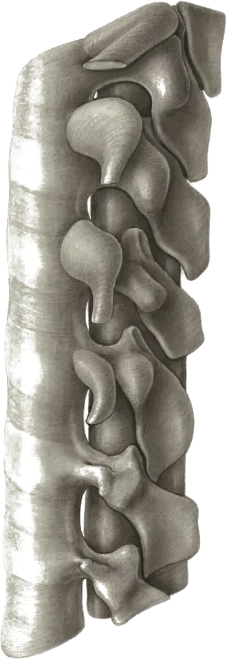
Most herniations occur at L4-5 and L5-S1 levels in the lower back, or C5-6 and C6-7 in the neck.
Overview
Contributing Factors
Link copiedDisc are strongly linked to specific loading patterns and movement mechanics. The most dangerous combination is forward bending with rotation while lifting - this creates asymmetric loading that can cause the disc's outer ring to fail. When you bend forward and twist at the same time, you create massive shearing forces through the disc that are particularly damaging to the posterior and posterolateral aspects where most herniations occur.
Morning activities are particularly risky because your discs absorb fluid overnight, making them larger and more vulnerable to injury in the first hour after waking. Something as simple as bending over to pick up a sock or reaching for a toothbrush can be the final straw if your disc is already compromised. This is why many people report their disc injury happened during a seemingly minor activity.
Prolonged sitting followed by sudden movement is another classic pattern. When you sit for extended periods, your discs experience increased pressure and your hip flexors tighten. When you suddenly stand and bend forward, you combine high disc pressure with poor movement mechanics from tight hips and weak glutes. Poor lifting technique compounds this - lifting with a rounded back, lifting away from your body, or lifting while seated all dramatically increase the forces through your discs and make herniation more likely.
Symptoms
Clinical Presentation
Link copiedPrimary Symptoms
Associated Symptoms
Typical pattern
Often sudden onset with bending or lifting. Specific movements consistently aggravate. May have periods of improvement and flare-ups.
Symptoms
Differential Diagnosis
Link copiedConditions with similar presentations:
Lumbar Radiculopathy from Foraminal Stenosis
Key differences: Older patient, gradual onset, leg symptoms worse with extension and standing rather than flexion or sitting. Imaging often shows bony foraminal narrowing rather than a discrete extrusion. typically runs opposite to the classic pattern.
Deep Gluteal Syndrome / Piriformis Involvement
Key differences: Deep buttock pain with referral into the posterior thigh, rarely below the knee, no clear back pain, and a normal neurological exam. Reproduced by prolonged sitting, seated stretch, or palpation of the deep gluteal space, not by spinal loading.
Sacroiliac Joint Pain
Key differences: One-sided pain near the PSIS, often pointed to with one finger. Pain rarely extends below the knee and does not follow a pattern. A cluster of provocation tests (distraction, compression, thigh thrust, FABER, Gaenslen) is more informative than any single test.
Facet-Mediated Pain with Somatic Referral
Key differences: Paraspinal pain aggravated by extension and rotation, with referral into the buttock or posterior thigh that stops at or above the knee. is negative, neurological exam is normal, and symptoms do not or peripheralize with repeated movement testing.
Hip Intra-articular Pathology
Key differences: Anterior groin or lateral hip pain reproduced by or FABER rather than loading. Limited and painful hip internal rotation is often the clearest finding. Symptoms track with weight-bearing activity rather than spinal posture or Valsalva.
Cauda Equina Syndrome
Key differences: Saddle numbness, urinary retention or incontinence, faecal incontinence, bilateral leg weakness, or rapidly progressive neurological deficit. Any of these flips the clinical picture from physiotherapy to an emergency department. I screen for this every visit in anyone with significant symptoms.
When to seek professional help
Research
Key Research & Evidence
Peer-reviewed studies supporting the treatment approach.
Study
The probability of spontaneous regression of lumbar herniated disc: a systematic review (Chiu et al., Clin Rehabil 2015)
Key findings
Spontaneous regression of conservatively managed lumbar herniations was common, with higher resorption rates for larger herniations: about 96% of sequestrations, 70% of extrusions, 41% of protrusions, and 13% of bulges showed regression
Clinical relevance
Supports conservative management approach
Research Database Expanding
Additional peer-reviewed studies are being reviewed and will be added to strengthen the evidence base for this condition.
Management
Evidence-Based Management
Treatment strategies with the strongest support in the current literature.
Primary approach
-based exercises are consistently the most reliable early intervention for disc-related symptoms and often bring meaningful relief within the first several weeks
Complementary
techniques reduce nerve sensitivity and improve mobility when combined with directional exercises that promote of symptoms
Prevention & long-term
Movement education focusing on spine-sparing strategies and graded loading can reduce the likelihood of recurrent disc episodes
Detailed management strategies
Position Management
Finding positions that reduce nerve pressure promotes healing
Important precautions
- Avoid positions that worsen leg/arm symptoms
Gradual Loading
Progressive return to activities allows adaptation
Important precautions
- Monitor neurological symptoms
Management
Treatment Techniques
Evidence-based manual therapy and intervention approaches.
Treatment approaches supported by current research and clinical guidelines
Recommended treatment approaches
Treatment approaches are individualized to each patient's needs and goals. All interventions require explicit informed consent, and treatment plans are collaboratively modified based on your preferences and response to care.
Pain Education & Self-Management
Understanding pain science to reduce fear and improve movement confidence alongside active rehabilitation.
Exercise Therapy
Personalized exercise programs designed to restore strength, flexibility, and function.
Joint Mobilization
Graded techniques to restore joint movement and reduce stiffness.
Postural Assessment & Movement Strategies
Analysis of posture and movement patterns to develop adaptable positioning strategies.
Post-Surgical Rehabilitation
Evidence-based recovery programs following surgery to restore function and strength.
Rehabilitation
A Typical Rehabilitation Progression
Three phases, from settling symptoms to returning to full activity.
Recovery from Disc Herniations is usually staged: calm the symptoms first, then rebuild the strength and capacity of the area, then return to your full activities. The three phases below show the kind of progression the evidence supports and that I commonly work through in clinic. They are here to show you what the road can look like, not to act as a personal program.
- Phase 1
Settling Symptoms and Finding a Directional Preference
Reduce nerve irritation and identify which positions and movements consistently move pain away from the leg and back toward the spine. testing is a core recommendation of the JOSPT Clinical Practice Guidelines on low back pain (George et al., 2021).
Examples, not a prescription
- Prone press-ups every 2 to 3 hours if extension centralizes symptoms, 10 repetitions per set
- Supported prone lying or prone on elbows for 3 to 5 minutes as a resting position if tolerated
- Gentle sliders in long sitting or supine, 10 to 15 slow repetitions, stopping short of any increase in leg symptoms
- Short walking bouts of 5 to 10 minutes several times per day, using posture cues that feel best
- Standing breaks every 20 to 30 minutes and a small roll for sitting
Ready to progress when
Leg symptoms consistently stay at or above the knee, rest pain is 2/10 or lower, sitting tolerance reaches 20 to 30 minutes, and the patient can identify at least one movement that reliably centralizes symptoms.
- Phase 2
Restoring Load Tolerance and Motor Control
Rebuild trunk and hip capacity so the spine tolerates daily loads again. Graded exercise and education outperform passive care in the Lancet Low Back Pain Series (Foster et al., 2018). Neural mobility work progresses from sliders toward tensioners as leg symptoms settle.
Examples, not a prescription
- Bird dog and dead bug, 2 to 3 sets of 8 to 10 per side, emphasizing a neutral spine
- Hip hinge patterning from a broomstick to a kettlebell deadlift, progressed by load rather than range
- Goblet squat to a box, 3 sets of 8 to 10, depth guided by symptom response
- Glute bridges and side-lying to address gluteal weakness common in this group
- Short-range sciatic nerve tensioners in supine or slump, 8 to 10 controlled repetitions
Ready to progress when
Loaded hinging and squatting with pain under 3/10, a full workday with normal sitting tolerated, and 30 minutes of walking without symptom spread into the leg.
- Phase 3
Return to Full Activity and Reducing Recurrence
Rebuild capacity for the specific demands the patient is returning to, whether lifting at work, running, or recreational sport. Heavier loading and higher-velocity tasks are layered in, with a simple maintenance plan to reduce the risk of another episode.
Examples, not a prescription
- Trap-bar or conventional deadlift built progressively from light loads, 3 to 4 sets of 5
- Front-loaded squats or split squats at working loads, matched to patient goals
- Farmer and suitcase carries for trunk and grip, 3 to 4 rounds of 30 to 40 metres
- Graded return to running or sport using walk-run intervals as the entry point
- Twice-weekly maintenance strengthening that the patient can sustain on their own
Ready to progress when
Full work, home, and recreational demands with minimal or no symptoms, confidence in self-managing minor flares, and a written maintenance plan the patient owns.
Management
Prognosis & Recovery
What outcomes and recovery factors typically look like.
Expected timeline
Most improve significantly within 6-12 weeks. Full recovery 3-6 months
Natural history
Most get smaller over time on imaging, and most people recover without surgery
Factors affecting recovery
Management
Measuring Progress
How to track the recovery arc week to week.
Day-to-day tracking
I track what changes day to day: pain interference with key tasks, movement quality during functional tests, and your confidence with daily activities
Assessment tools
Condition-specific questionnaires when useful (like the Oswestry for back pain or DASH for shoulder conditions)
Activity targets
One activity target that matches your goal - whether that's returning to sport, work tasks, or daily activities without limitation
Management
Frequently Asked Questions
Common concerns and answers about this condition.
Will my disc herniation heal on its own?
Will my disc herniation heal on its own?
Often, yes. Systematic reviews of serial MRI studies show that most shrink over time, with higher resorption rates for larger extrusions and sequestrations than for small bulges or protrusions. Symptom improvement usually precedes imaging change, which is why I track function rather than imaging week to week. The size of the herniation on your scan does not neatly predict how bad it feels, nor how quickly you will recover.
Do I need surgery for a herniated disc?
Do I need surgery for a herniated disc?
Most people do not. The SPORT trial (Weinstein et al., JAMA 2006) and its follow-up studies found that at 1 to 2 years, outcomes between surgery and structured conservative care for disc converge, though surgery can produce faster early relief. Surgery is typically reserved for progressive neurological loss, , or pain and disability that remain severe after 6 to 12 weeks of appropriate non-surgical management.
How long does recovery take?
How long does recovery take?
Most people see meaningful change within 4 to 6 weeks, and the majority recover within 3 months. Full resolution of residual symptoms can take 3 to 6 months, sometimes longer for larger . Recovery is rarely a straight line. I watch for , improved sitting tolerance, and return of strength rather than waiting for a perfect pain-free day.
My MRI says disc herniation. Does that mean I am damaged?
My MRI says disc herniation. Does that mean I am damaged?
Not in the way people usually fear. Jensen et al. (NEJM 1994) found disc bulges in 52% of pain-free adults and protrusions in 27%. Brinjikji et al. (AJNR 2015) reported disc in 37% of asymptomatic 20-year-olds and 96% of 80-year-olds. Imaging findings are common in people with no back pain at all. A on your scan is a finding to interpret alongside your symptoms, your physical exam, and your goals, not a verdict on your spine.
Can I still exercise with a disc herniation?
Can I still exercise with a disc herniation?
In most cases, yes, and staying active is linked to better outcomes than rest in both NICE NG59 and the Lancet Low Back Pain Series. The specifics matter. Early on, I usually pull back on deep forward bending under load, heavy rotational lifting, and long runs, while keeping walking, supported positions, and symptom-guided loading in the program. What you can do changes week to week as symptoms settle.
What about epidural steroid injections?
What about epidural steroid injections?
NICE NG59 supports considering an epidural of local anaesthetic and steroid for acute and severe sciatica. The evidence base suggests short-term pain relief with limited long-term benefit and no clear effect on the need for surgery. They can be useful when pain is blocking progress with rehab, but they work best as a bridge to active treatment rather than as a standalone plan.
Should I avoid bending forward for good?
Should I avoid bending forward for good?
No. Permanent avoidance tends to produce a fragile, fearful back rather than a resilient one. There are reasons to respect flexion early on, especially first thing in the morning when discs are more hydrated and vulnerable, and when combining bending with twisting and load. The end goal is to bend and load the spine confidently again, in stages your tissues can handle.
When should I get urgent medical care?
When should I get urgent medical care?
Loss of bladder or bowel control, numbness in the saddle area, rapidly progressing leg weakness, foot drop, or bilateral leg symptoms all warrant an emergency department visit. Severe unrelenting night pain, unexplained weight loss, or fever with back pain should be reviewed by a physician before starting physiotherapy.
Related Conditions
Conditions I commonly see alongside, or confused with, this one.
- Common co-occurrence
Sciatica
Disc herniation is the most common cause of sciatica and nerve root compression
- Common co-occurrence
Low Back Pain
Disc herniation is often the underlying pathology in mechanical low back pain
- Common co-occurrence
Degenerative Disc Disease
Disc degeneration often precedes and predisposes to herniation