Tarsal Tunnel Syndrome
Posterior tibial nerve compression, medial ankle numbness
Overview
The Science of Tarsal Tunnel Syndrome
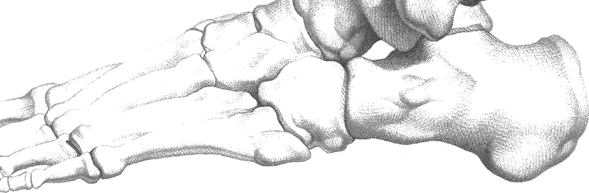
Link copiedTarsal tunnel syndrome results from compression of the posterior tibial nerve as it passes through the tarsal tunnel, a fibro-osseous space located behind the medial malleolus at the ankle. The tunnel is bounded by the flexor (laciniate ligament) superficially and the tibia, talus, and deep to the nerve.
The posterior tibial nerve carries sensory fibers to the plantar surface of the foot and motor fibers to most of the . Within or just distal to the tarsal tunnel, the nerve divides into medial and lateral plantar branches, and sometimes a medial calcaneal branch. Compression can affect the main trunk proximally or individual branches distally, creating variable symptom patterns.
Several mechanisms can cause nerve compression within this confined space. Space-occupying lesions such as ganglion cysts, lipomas, or varicose veins can directly compress neural structures. Inflammatory conditions like rheumatoid , , or chronic ankle can cause swelling that reduces the available space for the nerve.
Biomechanical factors contribute significantly to tarsal tunnel syndrome development. Excessive foot increases tension on the flexor retinaculum and can cause the nerve to bow around the medial malleolus, creating a functional compression. This explains why the condition often occurs in association with posterior tibial tendon dysfunction or flexible flatfoot deformity.
Chronic compression leads to intraneural edema, demyelination, and eventual axonal damage if left untreated. The progression from reversible conduction block to permanent nerve damage explains why early recognition and treatment are crucial for optimal outcomes.
Overview
Contributing Factors
Link copiedNormal posterior tibial nerve function requires unimpeded passage through the tarsal tunnel during all phases of gait. The tunnel dimensions change dynamically with foot and ankle position, with maximum space available in and , and minimum space in and .
During normal gait, the foot progresses through during loading response, followed by supination during push-off. This biomechanical sequence requires the tarsal tunnel structures to accommodate changing positions and loads. When excessive pronation occurs, several problems develop that can compromise nerve function.
Excessive pronation increases tension on the flexor as it attempts to maintain the tunnel's integrity against abnormal forces. This can create a bowstring effect where the nerve becomes compressed against the rigid medial malleolus. The prolonged pronated position also maintains the tunnel in its most confined configuration for longer periods during the gait cycle.
The posterior tibial tendon plays a crucial role in controlling pronation and maintaining the medial longitudinal arch. When this tendon becomes dysfunctional, excessive pronation results, directly affecting tarsal tunnel dimensions. This explains the frequent association between posterior tibial tendon dysfunction and tarsal tunnel syndrome.
Footwear and activity patterns influence significantly. Shoes with inadequate arch support allow excessive pronation, while high-heeled shoes can alter ankle positioning and tunnel dimensions. Prolonged standing or walking activities may exceed the nerve's tolerance for sustained compression, particularly in the presence of underlying biomechanical abnormalities.
Recovery requires not only addressing the acute nerve compression but also correcting the underlying biomechanical factors that contributed to the problem. This often involves orthotic management to control pronation and posterior tibial tendon rehabilitation to restore normal foot mechanics.
Symptoms
Clinical Presentation
Link copiedPrimary Symptoms
Associated Symptoms
Typical pattern
Symptoms typically follow a pattern of activity-related onset with improvement during rest periods. Night symptoms are characteristic and often more severe than daytime complaints. Bilateral involvement can occur, often related to systemic conditions or bilateral biomechanical abnormalities.
Symptoms
Differential Diagnosis
Link copiedConditions with similar presentations:
Plantar Fasciitis
Key differences: Pain primarily plantar heel, worse with first steps, lacks neurological symptoms
Posterior Tibial Tendon Dysfunction
Key differences: Medial arch pain with flatfoot deformity, weakness with single heel rise, lacks neurological symptoms
Morton's Neuroma
Key differences: location between , affected toes typically third and fourth
Diabetic Peripheral Neuropathy
Key differences: Bilateral symmetric distribution, burning feet syndrome, associated with diabetes mellitus
S1 Radiculopathy
Key differences: Back pain with radiation, affects entire leg, positive test
When to seek professional help
Research
Key Research & Evidence
Peer-reviewed studies supporting the treatment approach.
Finding
Surgical treatment is associated with excellent or good outcomes in 75.3% of cases across 32 studies
Research details
A 2024 scoping review (Haq et al., J Clin Orthop Trauma) of 32 studies reported that excellent or good results were seen in 75.3% of cases, with the remainder having fair or poor outcomes, though the authors noted evidence quality was low and outcome reporting varied across studies
Clinical relevance
While three-quarters of patients achieve good surgical outcomes, one-quarter experience suboptimal results with meaningful complication and recurrence rates, supporting conservative management as appropriate first-line treatment with surgery reserved for refractory cases after adequate trial of 4-6 months
Finding
17 of 32 surgical studies reported failed conservative treatment prior to surgery
Research details
The 2024 scoping review documented that 17 studies specifically reported failure of conservative treatment before proceeding to surgical nerve decompression, with factors influencing surgical outcomes including patient age, symptom duration, etiology, comorbidities, pre-treatment symptom severity, and nerve fibrosis, highlighting importance of patient selection and timing
Clinical relevance
Surgical decision-making requires consideration of multiple prognostic factors beyond simple failure of conservative care, with duration of conservative treatment, underlying etiology, and baseline symptom severity affecting likelihood of surgical success and supporting thorough conservative trial before operative intervention
Research Database Expanding
Additional peer-reviewed studies are being reviewed and will be added to strengthen the evidence base for this condition.
Management
Evidence-Based Management
Treatment strategies with the strongest support in the current literature.
Primary approach
Conservative treatment combining activity modification, , and biomechanical correction can achieve significant symptom improvement, particularly when initiated early in the course of symptoms
Complementary
Corticosteroid injections can provide temporary symptom relief but show variable long-term success rates and are typically reserved for cases not responding to conservative management
Prevention & long-term
Early recognition and treatment of underlying biomechanical factors and associated conditions can help prevent progression to chronic nerve compression
Detailed management strategies
Activity Pacing and Position Modification
Avoiding prolonged weight-bearing activities and positions that compress the nerve allows inflammation to subside and prevents further nerve irritation
Important precautions
- Complete activity cessation rarely necessary
- Modify rather than eliminate activities
- Use frequent position changes during prolonged activities
Supportive Footwear and Arch Support
Proper footwear with arch support helps control excessive and reduces stress on the tarsal tunnel, minimizing nerve compression during daily activities
Important precautions
- Professional fitting may be beneficial for optimal results
- Start with over-the-counter supports before progressing to custom options
- Ensure footwear accommodates any swelling
Gentle Neural Mobility Exercises
Specific exercises help maintain nerve mobility and prevent adhesion formation while promoting healing through improved blood flow
Important precautions
- Exercises should not reproduce or worsen neurological symptoms
- Stop immediately if numbness or tingling increases
- Perform gently and avoid aggressive stretching
Night Positioning and Sleep Optimization
Proper positioning during sleep reduces nerve compression and minimizes night symptoms that commonly disrupt sleep in tarsal tunnel syndrome
Important precautions
- Avoid sleeping on the affected side
- Use pillows to elevate the foot if helpful
- Consider loose-fitting socks to avoid constriction
Ice Application and Anti-inflammatory Management
Controlled ice application can reduce swelling within the tarsal tunnel and provide symptomatic relief, particularly during acute flare-ups
Important precautions
- Apply ice for 15-20 minutes maximum
- Use barrier between ice and skin
- Avoid ice if you have circulation problems or diabetes
Management
Treatment Techniques
Evidence-based manual therapy and intervention approaches.
Treatment approaches supported by current research and clinical guidelines
Management
Prognosis & Recovery
What outcomes and recovery factors typically look like.
Expected timeline
Early intervention within 6 months of symptom onset typically results in significant improvement within 8-12 weeks. Chronic cases may require 3-6 months of consistent treatment with more variable outcomes
Natural history
Early cases often respond well to conservative treatment with complete symptom resolution. Chronic compression can lead to permanent nerve damage with persistent numbness and weakness. Untreated cases may progress to irreversible functional limitations
Factors affecting recovery
Management
Measuring Progress
How to track the recovery arc week to week.
Day-to-day tracking
I monitor changes in your neurological symptoms including numbness and tingling, assess improvements in functional activities like walking and standing tolerance, evaluate sleep quality improvements, and track your ability to perform daily activities without nerve-related limitations
Assessment tools
Foot and Ankle Ability Measure (FAAM) for functional assessment, neuropathy-specific questionnaires for symptom tracking, and sleep quality assessments for monitoring night symptom improvements
Activity targets
Complete elimination of nerve-related symptoms with full return to desired activities, restoration of normal foot sensation and function, and prevention of symptom recurrence
Related Conditions
Conditions I commonly see alongside, or confused with, this one.
- Shares symptoms
Plantar Fasciitis & Heel Spurs
Both can cause medial arch and heel pain symptoms
- Anatomically related
Posterior Tibial Tendon Dysfunction
Both involve medial ankle structures; PTTD swelling can compress nerve
- Shares symptoms
Morton's Neuroma
Both cause foot numbness but in different nerve distributions