Patellar Tendinopathy (Jumper's Knee)
Patellar tendon pain common in jumping sports
Overview
The Science of Patellar Tendinopathy (Jumper's Knee)
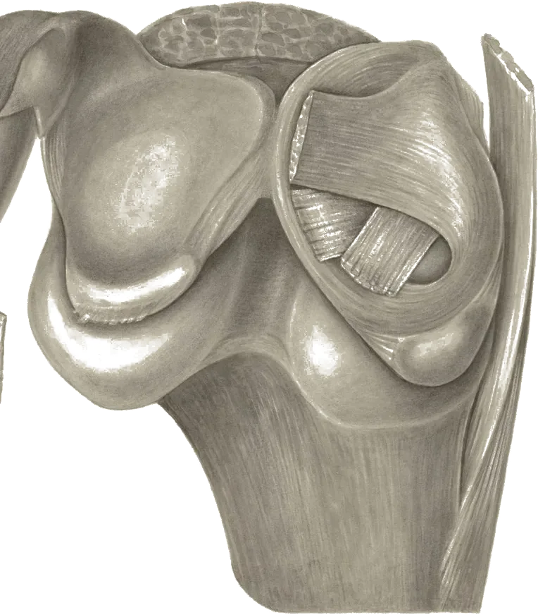
Link copiedPatellar represents a complex condition affecting the patellar tendon, predominantly at its attachment to the inferior pole of the patella. The condition involves progressive fiber disorganization and failed healing response rather than true inflammation, which fundamentally changes our approach to treatment.
The begins with repetitive microtrauma from jumping and landing activities that overwhelm the tendon's capacity to repair. This creates microscopic failures within the tendon structure, leading to alterations at the cellular level that undermine its mechanical properties. The normal parallel arrangement of type I collagen fibers becomes disrupted, replaced by areas of mucoid degeneration and increased ground substance that weakens the tendon's tensile strength.
At the cellular level, tenocytes undergo significant changes in response to repetitive loading. These cells alter their protein and enzyme production, increasing prostaglandin E2 and leukotriene B4, which contribute to the degenerative process. Matrix metalloproteinase activity increases, breaking down the extracellular matrix faster than it can be rebuilt. Simultaneously, vascular endothelial growth factor production leads to , bringing new blood vessels and nerve fibers into areas that are normally , contributing to pain sensation.
The tendon's appearance changes dramatically at the microscopic level. Instead of tightly packed, parallel collagen bundles, affected tendons show areas of fibrinoid necrosis, pseudocyst formation, and random collagen orientation. There's hypercellularity with atypical fibroblast proliferation and areas of cell death through apoptosis. This creates the characteristic thickened, painful tendon seen clinically, often described as having a "mucoid" appearance on imaging.
Importantly, this is primarily a degenerative rather than inflammatory condition. While acute inflammation may occur with initial injury, chronic patellar tendinopathy shows minimal inflammatory cells. This understanding has shifted treatment away from anti-inflammatory approaches toward loading programs that stimulate proper tendon remodeling and collagen synthesis.
Overview
Contributing Factors
Link copiedThe patellar tendon experiences extraordinary mechanical loads during jumping and landing activities that predispose athletes to . During the phase of landing from a jump, your quadriceps must generate high forces while lengthening to control knee flexion and decelerate your body's downward momentum. Research using force plates and motion capture shows that landing from a vertical jump creates patellar tendon forces reaching 6-8 times body weight, concentrated at the bone-tendon interface where most commonly develops.
Counter-movement jump performance serves as both a risk factor and biomechanical indicator for patellar tendinopathy development. A 2023 meta-analysis identified counter-movement jump height as a significant risk factor, representing explosive lower limb power through coordinated eccentric and muscle contractions. Athletes with higher jump heights generate greater tendon loading with each repetition. When you perform hundreds or thousands of jumps weekly in sports like volleyball or basketball, these accumulated high-magnitude loads can exceed the tendon's adaptive capacity, particularly during periods of rapid training volume increases.
Squatting mechanics dramatically influence patellar tendon loading magnitudes. Biomechanical studies demonstrate that performing squats on a 25-degree decline board maximizes patellar tendon strain compared to level-ground squats. During decline squats, your knees translate further forward over your toes, increasing the moment arm and requiring greater quadriceps force production. Research using ultrasound elastography shows significantly greater patellar tendon strain, smaller ankle and hip joint angles, and higher knee extensor muscle EMG amplitudes during decline squats. This explains why decline squat protocols effectively load the tendon therapeutically but can also contribute to overload if training volume isn't managed properly.
Sports involving high eccentric quadriceps loading create the greatest risk for patellar tendinopathy. Volleyball players performing spike approaches, basketball players landing from rebounds, long and high jumpers in track and field, long-distance runners absorbing repetitive impact forces, and skiers controlling descent all expose their patellar tendons to sustained eccentric loads. Studies tracking volleyball athletes show patellar tendinopathy prevalence rates of 40-50% in elite players, with rates correlating directly with years of competitive participation and weekly jump volume.
Hip and ankle significantly influence patellar tendon loading through effects. Reduced ankle range forces your knee to translate less far forward during squatting and landing, theoretically reducing patellar tendon moment arm. However, this compensation often increases hip flexion demands and alters landing mechanics in ways that can increase injury risk elsewhere. Research demonstrates that hip muscle weakness, particularly of the gluteus maximus and medius, associates with increased knee during landing, altering the line of quadriceps force application and creating asymmetrical patellar tendon loading patterns.
Training load progression rates critically affect whether tendon loading stimulates adaptation or causes breakdown. Your patellar tendon adapts to mechanical stress through increased synthesis and cross-linking, but this process requires 36-72 hours. When training volume or intensity increases too rapidly, you accumulate tendon microdamage faster than repair mechanisms can address it. Rapid increases in weekly jump volume relative to what the tendon has adapted to can elevate tendinopathy risk. Elite volleyball players transitioning from off-season to pre-season training demonstrate the highest injury rates during these rapid loading progressions.
Landing mechanics and knee flexion angles during ground contact determine peak patellar tendon forces. Stiff-legged landings with minimal knee flexion, common in fatigued athletes or those with pain-avoidance patterns, dramatically increase peak tendon loading. When you land with only 30-40 degrees of knee flexion versus 60-80 degrees, the reduced motion range means your quadriceps must generate higher forces over shorter time periods to dissipate the same kinetic energy. Biomechanical analysis shows that landing with reduced knee flexion can increase peak patellar tendon forces, explaining why fatigue-related technique breakdown contributes to injury risk.
Body mass significantly influences absolute patellar tendon loading. Each kilogram of body weight directly increases the gravitational force your tendon must resist during landing and deceleration. Research shows direct correlation between body mass index and patellar tendinopathy prevalence in jumping sports. Weight gain translates into additional peak tendon force during landing from a countermovement jump, representing a load increase that may exceed tendon adaptive capacity if weight gain occurs rapidly without proportional strength increases.
Symptoms
Clinical Presentation
Link copiedPrimary Symptoms
Associated Symptoms
Typical pattern
Patellar follows a predictable progression through distinct stages. Initially, pain occurs only after intense activity and doesn't affect performance. This progresses to pain during activity that may warm up but returns worse afterward. Advanced stages involve pain during daily activities and inability to participate in sports. The hallmark feature is load-related pain that increases predictably with energy storage activities like jumping. Athletes often describe being able to pinpoint the exact moment in training when pain will begin, such as after a specific number of jumps or at a particular point in their run.
Symptoms
Differential Diagnosis
Link copiedConditions with similar presentations:
Patellofemoral Pain Syndrome
Key differences: Diffuse pain behind or around the kneecap rather than focal tenderness at the inferior pole, aggravated by stairs, running, and prolonged sitting with knees bent, positive patellar compression test, and pain is generally not reproduced by single-leg decline squat at the tendon insertion.
Quadriceps Tendinopathy
Key differences: Tenderness at the superior pole of the patella rather than the inferior pole, pain with resisted knee extension and deep squatting, more common in weightlifters and middle-aged athletes than adolescent jumpers.
Hoffa's Fat Pad Impingement
Key differences: Pain and swelling in the infrapatellar region either side of the tendon, worse with terminal knee extension and prolonged standing, positive Hoffa's test with pain on active extension while the examiner compresses the fat pad.
Osgood-Schlatter Disease
Key differences: Focal pain and prominence at the tibial tubercle rather than the patellar inferior pole, typically ages 10 to 15 during growth spurts, visible bony enlargement at the tibial insertion.
Plica Syndrome
Key differences: Snapping or catching sensation along the medial border of the patella, palpable thickened band that is reproducibly tender, symptoms with repetitive flexion-extension rather than with pure loading tasks.
Meniscal Pathology
Key differences: Joint-line tenderness rather than tendon tenderness, mechanical symptoms including catching, locking, or a sensation of giving way, positive McMurray or Thessaly test, often a history of twisting injury.
Patellar Stress Fracture or Sinding-Larsen-Johansson
Key differences: Night pain, rest pain, and pain disproportionate to loading history, focal bony rather than tendon tenderness, recent rapid spike in training load, MRI showing bone oedema.
When to seek professional help
Research
Key Research & Evidence
Peer-reviewed studies supporting the treatment approach.
Finding
Evidence quality remains limited despite multiple treatment options
Research details
February 2024 review of meta-analyses revealed lack of high-quality evidence on optimal patellar tendinopathy treatments, though PRP and ESWT show promise. Evidence for eccentric exercise efficacy remains unclear due to inconclusive findings across studies
Clinical relevance
Clinical decision-making requires individualization based on patient presentation and response to trial interventions rather than assuming single protocol superiority given evidence limitations
Finding
Landing forces reach 6-8 times body weight in jumpers
Research details
Research using force plates and inverse-dynamics modelling shows landing from a vertical jump generates high patellar tendon forces, with estimates in the range of roughly 4-5 times body weight, concentrated at the bone-tendon interface. A 2018 systematic review and meta-analysis (Sprague et al., British Journal of Sports Medicine) examined modifiable risk factors for patellar tendinopathy and found only limited or conflicting evidence that greater counter-movement jump height is associated with developing the condition in athletes
Clinical relevance
Understanding high mechanical loads informs both prevention through landing technique optimization and rehabilitation progression matching tendon load tolerance capacity
Research Database Expanding
Additional peer-reviewed studies are being reviewed and will be added to strengthen the evidence base for this condition.
Management
Evidence-Based Management
Treatment strategies with the strongest support in the current literature.
Primary approach
Progressive tendon loading combining , heavy slow resistance, or moderate resistance training shows favourable outcomes compared with alone according to 2024 systematic reviews
Complementary
In-season protocols provide immediate pain relief allowing continued sport participation while longer-term heavy slow resistance programs build tendon capacity during off-season periods
Prevention & long-term
Gradual training load progression combined with landing technique and calf flexibility work can meaningfully reduce patellar incidence in jumping athletes
Detailed management strategies
Isometric Wall Sit or Spanish Squat Hold
Provides immediate pain relief through cortical inhibition while beginning to load the tendon in a controlled manner. Holding for 45 seconds at 70% effort stimulates tendon adaptation
Important precautions
- Pain should not exceed 5/10 during exercise
- Stop if pain remains elevated 24 hours post-exercise
- Maintain proper knee alignment over toes
Progressive Loading Using Decline Squat
The decline position loads the patellar tendon more than a level-surface squat, giving a useful therapeutic stimulus while being easily controlled
Important precautions
- Start with body weight only before adding resistance
- Use slow, controlled tempo (3 seconds down, 3 up)
- Some pain during exercise is acceptable but should settle within 24 hours
Calf and Quadriceps Flexibility Program
Reduced ankle shifts more landing load onto the patellar tendon. Daily calf stretching and foam rolling reduce this mechanical disadvantage
Important precautions
- Stretch after warming up, not when cold
- Hold stretches for 30-45 seconds
- Avoid aggressive stretching that provokes tendon pain
Modified Training Load Management
Continuing modified training maintains fitness and tissue capacity while allowing healing. Reducing jumping volume by 50% while maintaining other training often allows sport continuation
Important precautions
- Monitor total weekly jumping/landing count
- Implement hard/easy day training pattern
- Increase load by maximum 10% per week when returning
Energy Storage Exercise Progression
Gradual return to jumping and landing retrains the tendon's spring-like function. Starting with small hops and progressing to sport-specific movements ensures complete rehabilitation
Important precautions
- Only begin when pain-free with heavy resistance exercises
- Start with bilateral before unilateral activities
- Quality over quantity - maintain good landing mechanics
Management
Treatment Techniques
Evidence-based manual therapy and intervention approaches.
Treatment approaches supported by current research and clinical guidelines
Recommended treatment approaches
Treatment approaches are individualized to each patient's needs and goals. All interventions require explicit informed consent, and treatment plans are collaboratively modified based on your preferences and response to care.
Sports Rehabilitation & Return to Sport
Evidence-based recovery programs for athletes to safely return to sport after injury.
Dry Needling
Precise needle therapy targeting trigger points for effective pain relief and improved muscle function.
Exercise Therapy
Personalized exercise programs designed to restore strength, flexibility, and function.
Soft Tissue & Myofascial Therapy
Targeted hands-on techniques to address muscle tension, pain, and movement restrictions.
IASTM (Instrument Assisted Soft Tissue Mobilization)
Instrument-assisted techniques to address soft tissue restrictions and pain.
Rehabilitation
A Typical Rehabilitation Progression
Three phases, from settling symptoms to returning to full activity.
Recovery from Patellar Tendinopathy is usually staged: calm the symptoms first, then rebuild the strength and capacity of the area, then return to your full activities. The three phases below show the kind of progression the evidence supports and that I commonly work through in clinic. They are here to show you what the road can look like, not to act as a personal program.
- Phase 1
Isometric Loading and Pain Control (Weeks 1 to 4)
Reduce pain enough to train, start loading the tendon in a predictable way, and establish the 24-hour symptom response rule as the guide for progression. provide short-term analgesia through cortical inhibition, shown in Rio and colleagues' 2015 work in the British Journal of Sports Medicine, which makes them ideal for in-season athletes.
Examples, not a prescription
- Spanish squat isometric hold with a band around the knees, 5 sets of 45 seconds at roughly 70 percent effort, once daily
- Leg extension isometric hold at 60 degrees knee flexion, 5 sets of 45 seconds
- Wall sit with feet slightly forward, 5 sets of 45 seconds, progressed as tolerated
- Single-leg calf raise, 3 sets of 12, addressing calf capacity that offloads the tendon
- Modified sport volume, typically reducing jumping and sprinting by around 50 percent while preserving fitness work
Ready to progress when
Pain during Spanish squat hold at or below 3 out of 10, morning stiffness lasting under 10 minutes, and no more than mild symptom rise 24 hours after sessions for 7 consecutive days.
- Phase 2
Heavy Slow Resistance Training (Weeks 4 to 12)
Build tendon structural capacity using the heavy slow resistance protocol developed by Kongsgaard and colleagues in the Scandinavian Journal of Medicine and Science in Sports, which shows outcomes equivalent to programs with substantially better adherence. Tempo is 3 seconds down and 3 seconds up under heavier loads. Some pain during loading, up to 5 out of 10, is acceptable provided it settles within 24 hours.
Examples, not a prescription
- Heavy slow resistance leg press, 4 sets of 6 to 15 reps, 3 times per week on non-consecutive days, progressing from 15RM to 6RM over the block
- Heavy slow resistance barbell back squat or goblet squat, same tempo and set structure
- Decline squat on a 25-degree board with added load, 4 sets of 8 to 10 reps, twice per week
- Single-leg press, 3 sets of 8 to 10 reps, to address side-to-side asymmetry
- Continued calf and strengthening, 3 sets of 10 to 12 reps
Ready to progress when
score increased by at least 13 points from baseline, single-leg decline squat pain 3 out of 10 or less with full body weight, and heavy slow resistance sessions tolerated without 24-hour symptom flare for at least 2 weeks.
- Phase 3
Energy Storage and Return to Sport (Months 3 to 6)
Re-introduce the spring-like, stretch-shortening function of the tendon. This follows the Malliaras and colleagues staged loading framework: only progress to once heavy slow resistance is comfortable. Sport-specific demands, such as cutting, repeated jumping, and landing mechanics, are rebuilt gradually.
Examples, not a prescription
- Countermovement jumps, starting at 2 sets of 5 on soft surface, progressing to 4 sets of 10 on firm ground
- Drop jumps from a low box, progressing height only when landing mechanics stay clean
- Bounding and single-leg hops in straight lines, then with change of direction
- Sport-specific drills, such as approach jumps for volleyball or rebounding for basketball, in graded volume
- Return to full training using the 10 percent rule, increasing weekly jumping or sprinting volume by no more than 10 percent
Ready to progress when
VISA-P score above 80, pain 2 out of 10 or less during sport-specific tasks, symmetrical single-leg hop distance within 10 percent of the unaffected side, and two consecutive weeks of full sport training without a 24-hour flare.
Management
Prognosis & Recovery
What outcomes and recovery factors typically look like.
Expected timeline
Most athletes experience significant improvement within 12 weeks of appropriate loading programs, with many returning to sport over the following months, though recovery timelines vary considerably. However, complete tendon remodeling continues for 6-12 months. In-season athletes can often continue playing with modified training while undergoing rehabilitation
Natural history
Without appropriate treatment, around one-third of athletes are unable to return to sport within six months (Cook et al. 1997), and roughly half ultimately retire because of persistent symptoms (Kettunen et al. 2002). With evidence-based loading programs, many athletes return to their previous level of sport, though patience is required as biological healing cannot be rushed
Factors affecting recovery
Management
Measuring Progress
How to track the recovery arc week to week.
Day-to-day tracking
I track pain levels using the numeric rating scale (0-10) during specific loading activities like single-leg decline squat and jumping. We monitor the 24-hour symptom response to ensure appropriate loading. Weekly assessment of maximum quadriceps strength provides objective strength measures
Assessment tools
The Victorian Institute of Sport Assessment-Patella (VISA-P) questionnaire is our primary outcome measure, with scores below 80 indicating dysfunction. A 13-point change represents clinically meaningful improvement. We reassess every 4 weeks to track progress
Activity targets
Progressive return through defined stages: pain-free daily activities, then gym-based loading, followed by straight-line running, then sport-specific drills, and finally unrestricted sport participation. Each stage requires symptoms to remain stable for one week before progression
Management
Frequently Asked Questions
Common concerns and answers about this condition.
How long does patellar tendinopathy take to heal?
How long does patellar tendinopathy take to heal?
Most people notice meaningful improvement within 12 weeks of a structured loading program, but full tendon remodelling continues for 6 to 12 months. Longer symptom duration before starting rehab consistently predicts slower recovery. Athletes who start a loading program within a few months of symptom onset generally do better than those whose symptoms have been present for over a year. Consistency with loading matters more than intensity, and realistic expectations up front help patients stay committed during the slow biological timeline.
Should I stop running or jumping if I have jumper's knee?
Should I stop running or jumping if I have jumper's knee?
Complete rest is rarely the answer. Tendons need load to remodel, and prolonged rest often weakens them further. The usual approach is modifying volume rather than stopping: reducing jumping and high-speed running by around 50% and adding heavy slow resistance work often allows continued sport participation. The 24-hour rule guides dosage. If pain during activity stays under 5/10 and settles within 24 hours, the load was acceptable.
Does rest alone help patellar tendinopathy?
Does rest alone help patellar tendinopathy?
Rest can reduce pain short-term, but it does not stimulate the structural adaptation the tendon needs. The consistent message across recent systematic reviews and clinical guidance is that progressive loading is the primary treatment. Patients often cycle through weeks of rest, return to sport, and find symptoms come back within days. Loading exercises rebuild the tendon's capacity so it can handle sport demands again.
Are cortisone injections safe for patellar tendinopathy?
Are cortisone injections safe for patellar tendinopathy?
I advise caution. An injection can reduce pain short-term, but the Coombes et al. Lancet 2010 systematic review showed that for , corticosteroid injection produces worse outcomes at intermediate and long term compared with other interventions, including exercise. Corticosteroid can also weaken tendon structure. I use cortisone conversations as a chance to explain why loading, not suppression of symptoms, is the route back to sport.
When does patellar tendinopathy need surgery?
When does patellar tendinopathy need surgery?
Surgery is uncommon, and reserved for patients who have completed at least six months of well-structured loading rehabilitation without meaningful progress. Even then, surgical outcomes are not dramatically better than continued conservative care in most studies. Before considering surgery, I review whether the loading program was truly progressive, whether the hip, ankle, and calf were addressed, and whether sleep, training load, and nutrition have been optimised.
Can I keep playing sport during the season while rehabbing jumper's knee?
Can I keep playing sport during the season while rehabbing jumper's knee?
Often yes. Rio et al. (BJSM 2015) showed that holds (Spanish squats or wall sits at around 70% effort for 5 sets of 45 seconds) produce meaningful pain relief for roughly 45 minutes afterward, which is useful before competition or training. A reasonable in-season plan is isometrics plus modified jumping volume, then building heavy slow resistance work more aggressively in the off-season.
What is the difference between patellar tendinopathy and patellofemoral pain?
What is the difference between patellar tendinopathy and patellofemoral pain?
Patellar causes focal pain and tenderness at the bottom tip of the kneecap where the tendon attaches, and it behaves predictably with loading activities like jumping or decline squats. pain is more diffuse, sits behind or around the kneecap, and is aggravated by stairs, running, and prolonged sitting with bent knees. The distinction matters because the rehabilitation programs differ significantly in focus and progression.
Why does my knee hurt more the day after exercise?
Why does my knee hurt more the day after exercise?
Tendons respond to load over 24 to 48 hours, so the morning after heavier activity is often when symptoms peak. If pain rises above baseline and stays elevated for more than 24 hours, the previous session was too much for current tendon capacity. That is real-time feedback, not a reason to stop. Adjust the next session's load down and continue the program.
Related Conditions
Conditions I commonly see alongside, or confused with, this one.
- Anatomically related
Knee Pain
Both affect patellofemoral region; share many biomechanical risk factors
- Biomechanically linked
ACL Injuries
Both common in jumping sports; altered landing mechanics affect both structures
- Biomechanically linked
Hip Osteoarthritis
Hip stiffness can alter knee mechanics and increase patellar tendon stress
Commonly confused with
Side-by-side comparisons for patterns that often get mistaken for patellar tendinopathy (jumper's knee).
Get Expert Treatment
Professional physiotherapy for patellar tendinopathy (jumper's knee)