
Midfoot injury and rehabilitation
Lisfranc Injury: Midfoot Stability and Recovery
A Lisfranc injury affects the joints and supporting ligaments where the long bones of the foot meet the midfoot. It can involve a ligament tear, a fracture or joints moving out of alignment. The first treatment question is whether this part of the foot remains stable under load.
AAOS OrthoInfo (accessed), 2026When to see a doctor firstThe midfoot supports the arch and push-off
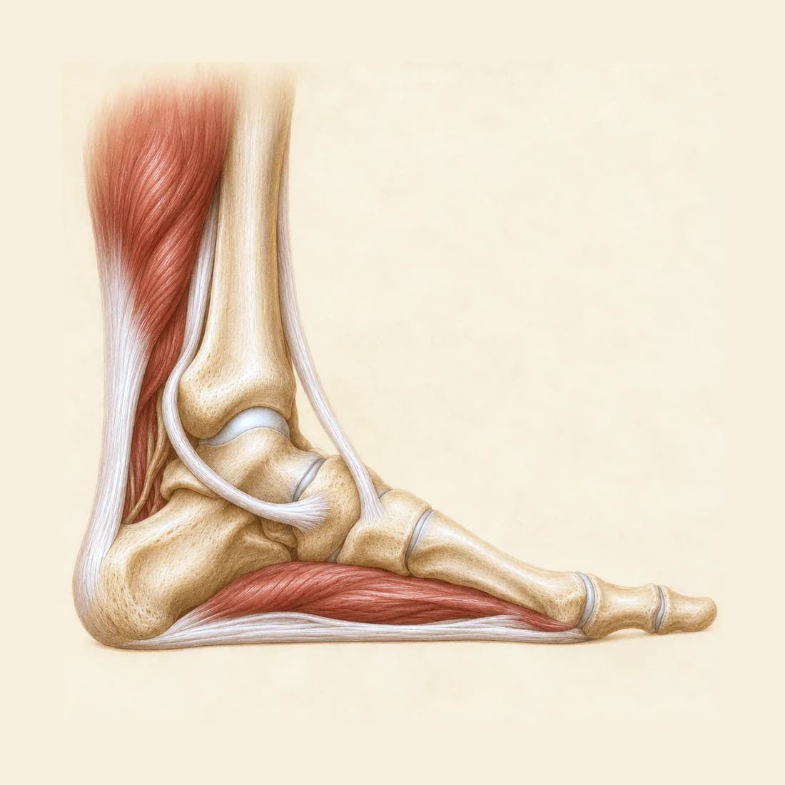
The metatarsals are the long bones leading toward the toes. Their bases meet the smaller midfoot bones at the tarsometatarsal joints. Ligaments hold this region together so it can support the arch and transfer force as you push off the ground. AAOS OrthoInfo (accessed), 2026
A Lisfranc injury can happen in a fall, a crush injury or a twist through the foot. The event can seem modest. Pain in this region after a twist deserves attention even when the injury was initially described as a sprained ankle. Semelsberger et al., 2026
Pain in the middle of the foot changes the assessment
Typical concerns include swelling over the middle of the foot, pain when standing or pushing off and bruising on the sole. Bruising underneath the foot is particularly concerning after an injury. These findings prompt assessment; you do not need to have every one of them. Semelsberger et al., 2026
Do not try to prove the foot is stable by hopping, rising onto the toes or twisting it yourself. A clinician chooses the examination and imaging. If the midfoot remains painful or swollen after an initial assessment or normal X-ray, arrange another review rather than continuing sport. AAOS OrthoInfo (accessed), 2026; Semelsberger et al., 2026
This differs from gradual bone stress pain after repeated loading and from a typical outer ankle sprain. The location and onset help decide which assessment is needed.
When to see a doctor first
Go to emergency
A foot that is visibly out of shape, an open wound over the injury, or a foot that is cold, pale or numb after trauma
Go to emergency now.
Get seen today
Midfoot pain and swelling after an injury, especially with bruising under the foot or difficulty putting weight on it
Keep weight off the painful foot and get medical assessment today through urgent care or your family doctor. A Lisfranc injury needs assessment before starting sprain exercises.
X-ray, CT and MRI answer different questions
The medical team decides which images are needed and how they can be taken safely. Standing, or weight-bearing, views can reveal widening that was not visible with the foot unloaded. Do not force yourself to stand for a home comparison or practise walking to test the injury. AAOS OrthoInfo (accessed), 2026
- Weight-bearing X-rays
- Show alignment under load and may be compared with the other foot. A normal initial film, particularly one taken without weight through the foot, can leave a suspected injury unresolved. Semelsberger et al., 2026
- CT
- Shows the bones, small fractures and joint alignment in more detail. Weight-bearing CT, where available and appropriate, can add information about alignment under load. Semelsberger et al., 2026
- MRI
- Shows the supporting ligaments and other soft tissues. It can help when a ligament injury remains in question after the initial assessment. Semelsberger et al., 2026
Stability guides protection and the surgical discussion
An injury confirmed to be stable can be managed without surgery, with an agreed protection plan and follow-up. A boot or cast and limits on weight bearing may be needed. Repeat examination or imaging checks that alignment is maintained as recovery progresses. Stødle et al., 2022
Displacement means the bones have shifted; diastasis means widening between structures that should sit together. Instability means the joints do not remain adequately supported. Those findings lead to an orthopaedic discussion about restoring alignment and stability. Semelsberger et al., 2026
Open reduction and internal fixation, or ORIF, realigns the injured joints and holds them with hardware. Arthrodesis, or fusion, joins selected injured joints together. Ask which joints are involved, why that operation is proposed and whether later hardware removal is expected. AAOS OrthoInfo (accessed), 2026
What the treatment studies tell us
A prospective cohort followed 26 people with CT-detected injuries that had not shifted and were confirmed stable with a clinician-performed stress test under fluoroscopy, a moving X-ray. They used a non-weight-bearing cast for six weeks in that study. Outcomes were excellent at a median follow-up of 55 months, and the foot-function score continued improving after the first year. The findings concern injuries whose stability had been checked. Stødle et al., 2022
The athlete consensus allows earlier weight bearing for selected stable injuries with close monitoring. That differs from the cohort’s protection plan. The assessment and the treating team’s instructions determine which approach fits your injury. Semelsberger et al., 2026; Stødle et al., 2022
A randomized trial of 43 displaced injuries compared fixation with primary fusion. It did not find clear evidence of a difference in the main foot-and-ankle outcome at two years, but recruitment fell short of the planned sample. Both remain options; this small trial does not settle the choice for every injury. Ponkilainen et al., 2024
Rehabilitation begins within the protection plan
Start with clear instructions about weight bearing, the boot or cast, permitted movement and the next review. After surgery, the operation and healing seen at follow-up guide those limits. Feeling less pain does not by itself change the written precautions. AAOS OrthoInfo (accessed), 2026
As loading is permitted, the plan connects movement and strength work with the walking you need. For example, a review may consider use of crutches, gait, swelling after an outing and the next practical task at home or work. Those observations help choose the next step within the agreed limits.
The post-surgical rehabilitation page explains how a procedure’s instructions shape exercise. The rehabilitation-progress guide helps you describe changes in activity and symptoms between appointments.
Walking, a full workday and sport need different preparation
Returning to modified participation, returning to sport and recovering previous performance are different stages. Comfortable daily walking leaves other demands to rebuild, such as longer time on your feet, repeated push-off, running or changes of direction. Ardern et al., 2016
Once those activities are permitted, preparation should reflect the activity you want back. The return-to-training guide explains how exercise, testing, confidence and practice fit together. The next review should identify what you can progress yourself and what still needs clearance. Ardern et al., 2016
Persistent midfoot pain, stiffness or swelling deserves review, particularly when walking is becoming harder. Follow-up may need to revisit the joints, healing or post-traumatic arthritis as well as the exercise dose. Semelsberger et al., 2026
Questions
Can I have a Lisfranc injury without a fracture?
Yes. The injury can involve the supporting ligaments without an obvious fracture. The assessment still needs to establish whether the midfoot remains stable, which can require further imaging when the first X-ray is unrevealing. Semelsberger et al., 2026
Does “nondisplaced” mean I can start walking normally?
Get the weight-bearing plan confirmed. Nondisplaced describes the position seen on the images. The successful non-surgical cohort also checked stability and used a protection plan with follow-up. The report wording alone is not permission to remove a boot or stop using crutches. Stødle et al., 2022
How long until I can run or return to sport?
The injury, treatment, healing and demands of the sport shape that answer. The athlete consensus concerns selected high-performance athletes and includes expert recommendations based on limited evidence. Its shorter return times should not be applied to an unstable injury or a healing fixation or fusion. Agree the next permitted activity and the review needed before running. Semelsberger et al., 2026
Sources and further reading
- AAOS OrthoInfo (accessed) · 2026 · Patient information
Lisfranc (Midfoot) Injury
Anatomy, recognition, imaging and treatment descriptions from the American Academy of Orthopaedic Surgeons. Accessed 16 November 2026; the page does not display a publication date.
- Semelsberger et al. · 2026 · Knee Surgery, Sports Traumatology, Arthroscopy
Diagnostic evaluation and nonoperative management of Lisfranc injuries in athletes.
International expert consensus and a systematic review focused on high-performance athletes. Supports assessment of stability, weight-bearing imaging and monitored non-surgical care for selected stable injuries. Its return times are not a general timetable for every Lisfranc injury.
- Stødle et al. · 2022 · Foot and Ankle Surgery
Outcome after nonoperative treatment of stable Lisfranc injuries. A prospective cohort study.
Followed 26 CT-detected, nondisplaced injuries confirmed stable by fluoroscopic stress testing. Non-surgical care included a protected period and follow-up imaging. Good results in this selected group do not establish stability from the word nondisplaced alone.
- Ponkilainen et al. · 2024 · Foot & Ankle International
Open Reduction Internal Fixation vs Primary Arthrodesis for Displaced Lisfranc Injuries: A Multicenter Randomized Controlled Trial.
Randomized 43 displaced injuries to fixation or primary fusion. The main outcome did not clearly differ at two years. The study did not reach its planned sample size and remained underpowered.
- Ardern et al. · 2016 · British Journal of Sports Medicine
2016 Consensus statement on return to sport from the First World Congress in Sports Physical Therapy, Bern.
Distinguishes return to participation, return to sport and return to performance. It presents return as a shared process informed by the injury, the activity and the athlete’s goals.