
Understanding pain and movement
The Rotator Cuff: Muscles, Tendons and Movement
The rotator cuff is a group of four muscles and their tendons around the shoulder joint. They connect the shoulder blade to the upper arm bone and work together to support the joint as the arm moves. The cuff includes supraspinatus, infraspinatus, teres minor and subscapularis.
La Rose et al., 2026; Cavanaugh et al., 2024Where the rotator cuff is
The shoulder joint joins the rounded head of the upper arm bone, or humerus, to the shallow glenoid socket on the shoulder blade, or scapula. This arrangement allows considerable movement. The capsule, labrum and ligaments contribute to stability alongside the surrounding muscles. La Rose et al., 2026
The cuff muscles begin on different surfaces of the scapula. Their tendons approach the humeral head from above, behind and in front. The muscles and tendons together form the cuff around the joint; a tendon is the part that transmits the muscle’s force into the bone. La Rose et al., 2026; Cavanaugh et al., 2024
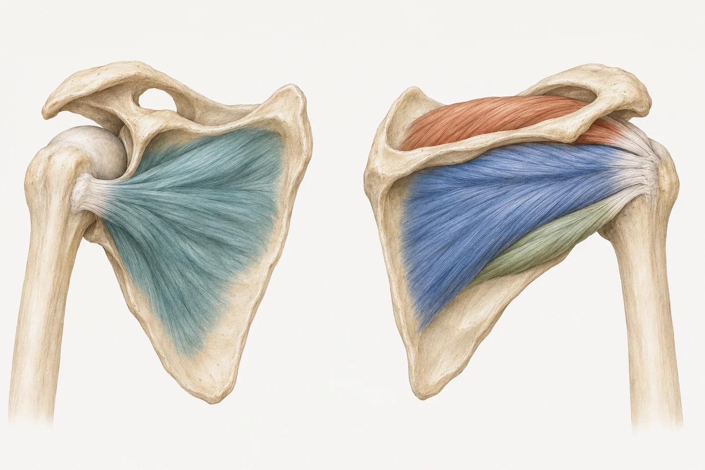
A teaching illustration with the outer shoulder muscles removed to show the cuff. Choosing a muscle keeps its broad region in colour; the regions are not measurements of tendon attachment sites.
- Supraspinatus Back view
- Above the spine of the shoulder blade. Contributes to lifting the arm and supporting the joint.
- Infraspinatus Back view
- Below the spine of the shoulder blade. Contributes to outward rotation and joint support.
- Teres minor Back view
- Along the outer back border of the shoulder blade. Contributes to outward rotation and joint support.
- Subscapularis Front view
- On the front surface of the shoulder blade, facing the rib cage. Contributes to inward rotation and joint support.
The four muscles
The names identify distinct muscles, but their work overlaps. Each contributes to shoulder control as well as movement. The contribution depends on the position of the arm and the task, so the actions below are a useful starting point rather than four isolated jobs. La Rose et al., 2026; Cavanaugh et al., 2024
- Supraspinatus
- Occupies the upper part of the back of the scapula, above its spine. Its tendon passes towards the top of the humerus. It contributes to lifting the arm and supporting the shoulder joint. Cavanaugh et al., 2024
- Infraspinatus
- Occupies much of the back of the scapula below its spine. Its tendon attaches to the back and upper side of the humerus. It contributes to outward rotation and joint support. Cavanaugh et al., 2024
- Teres minor
- Runs along the outer back border of the scapula towards the humerus. It contributes to outward rotation and works with the posterior cuff to support the joint. Cavanaugh et al., 2024
- Subscapularis
- Covers the front surface of the scapula, facing the rib cage, and attaches to the lesser tuberosity at the front of the humerus. It contributes to inward rotation and joint support. Cavanaugh et al., 2024
How movement and stability work together
Lifting or rotating the arm involves coordinated activity across the shoulder. The cuff contributes forces around the humeral head while other muscles help move the arm and shoulder blade. Stability is an active part of movement, rather than a separate task performed only when the arm is still. La Rose et al., 2026; Cavanaugh et al., 2024
The deltoid is a large shoulder muscle, but it is not one of the four cuff muscles. The long head of the biceps also contributes to the shoulder’s dynamic stabilisation and is considered alongside the cuff in the anatomy of the joint. La Rose et al., 2026; Cavanaugh et al., 2024
An outward-rotation exercise and an overhead task therefore involve related but different demands. Anatomy helps explain what an exercise asks of the shoulder; it does not establish which single muscle is responsible for a person’s pain. Cavanaugh et al., 2024; Desmeules et al., 2025
Muscle subregions and changing leverage
Cuff muscles contain subregions with different fibre arrangements, attachments and activation patterns. Their moment arms, which describe mechanical leverage around a joint, change with arm position. A muscle’s contribution is more complex than one movement written beside its name. Cavanaugh et al., 2024
Much of the detailed functional research examines specific positions or tasks, including throwing. Those findings explain mechanisms but should not be translated directly into a personalised diagnosis or exercise programme from the illustration alone. Cavanaugh et al., 2024
Muscle, tendon and the surrounding structures
The fleshy part of a cuff muscle produces force. The tendon transmits that force to the humerus. Reports may name the muscle when describing the tendon, which is why a scan can say supraspinatus tendinosis even though the finding concerns the tendon at the shoulder. La Rose et al., 2026; Cavanaugh et al., 2024; Goerl, 2020
The shoulder is supported by several structures, including the capsule, labrum, ligaments and muscles. Their contributions overlap. The cuff’s anatomy is one part of understanding shoulder movement and should be considered alongside the rest of the joint. La Rose et al., 2026
For the distinction between a clinical tendon problem and a tissue description, read tendinitis, tendinosis and tendinopathy. The supraspinatus guide explains the scan terminology used for this part of the cuff. Goerl, 2020; Gehwolf et al., 2025
When the anatomy appears on a report
A shoulder report may describe tendinopathy, calcification or a partial-thickness tear. These findings are included within the scope of the current rotator cuff tendinopathy clinical practice guideline, alongside assessment, rehabilitation and return to activity. Desmeules et al., 2025
A full-thickness tear requires its own clinical discussion. The tendinopathy guideline includes partial-thickness tears; it should not be used as though every recommendation necessarily applies to every full-thickness tear. The rotator cuff injury page covers the broader clinical problem. Desmeules et al., 2025
The anatomical name locates the structure. It does not by itself establish why you hurt or how much function is affected. The assessment combines the report with the history, movement and functional findings. Desmeules et al., 2025; Raja et al., 2020
What anatomy contributes to rehabilitation
Understanding the cuff can make the purpose of an exercise clearer. Rotation, lifting and shoulder control place different demands on the muscle group. The exercise needs to fit the clinical problem and the function you want to recover, rather than simply matching the muscle named on a scan. Cavanaugh et al., 2024; Desmeules et al., 2025
The current guideline addresses assessment, nonsurgical care, rehabilitation and return to function or sport for adults with rotator cuff tendinopathy. A drawing cannot supply the examination findings needed to apply that guidance to an individual shoulder. Desmeules et al., 2025
For a painful shoulder, start with the shoulder pain guide. Its medical-help section explains what needs assessment first, including symptoms following an injury. The anatomy here provides the background for those more specific explanations. Desmeules et al., 2025
Questions
Is the rotator cuff one muscle?
It includes four muscles and their tendons: supraspinatus, infraspinatus, teres minor and subscapularis. They work together around the shoulder joint. La Rose et al., 2026
Is the biceps part of the rotator cuff?
The biceps is not one of the four cuff muscles. Its long head contributes to dynamic shoulder stability and is an important neighbouring structure. La Rose et al., 2026
Which cuff muscles rotate the arm?
Infraspinatus and teres minor contribute to outward rotation, while subscapularis contributes to inward rotation. The cuff muscles also support the joint, and their contributions change with arm position. Cavanaugh et al., 2024
Can one exercise train every part of the cuff in the same way?
The cuff has several muscles and functional subregions, with contributions that change by task and arm position. Exercise selection should account for those demands alongside the clinical assessment. Cavanaugh et al., 2024; Desmeules et al., 2025
Sources and further reading
- La Rose et al. · 2026 · StatPearls
Anatomy, Rotator Cuff.
Describes the four cuff muscles and their role among the shoulder’s static and dynamic stabilisers.
- Cavanaugh et al. · 2024 · Sports
Subregions of the Rotator Cuff Muscles Present Distinct Anatomy, Biomechanics, and Function.
Describes cuff attachments, muscle subregions and how their contributions change with the task and arm position.
- Desmeules et al. · 2025 · JOSPT
Rotator Cuff Tendinopathy Diagnosis, Nonsurgical Medical Care, and Rehabilitation: A Clinical Practice Guideline.
Covers assessment, nonsurgical care and rehabilitation of adults with rotator cuff tendinopathy, including calcific tendinopathy and partial-thickness tears.
- Goerl · 2020 · The Journal of Family Practice
Getting tendinopathy treatment (and terminology) right.
Tendinopathy, tendinitis, tendinosis and paratenonitis describe different concepts rather than interchangeable tissue diagnoses.
- Gehwolf et al. · 2025 · Advanced Science
Tendinopathy: The Interplay between Mechanical Stress, Inflammation, and Vascularity.
Reviews interacting mechanical, inflammatory and vascular processes. Structural changes and pain do not have a simple one-to-one relationship.
- Raja et al. · 2020 · Pain
The revised International Association for the Study of Pain definition of pain: concepts, challenges, and compromises.
Defines pain as an experience and distinguishes it from nociception. Biological, psychological and social factors influence that experience.