
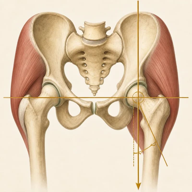
What Your Hip MRI or X-ray Report Means
A hip X-ray or MRI report lists what the pictures show, one structure at a time, in radiology shorthand. This page gathers the phrases that appear most often, gives each a short plain-language meaning, and links to its full guide. A report describes the picture. It does not say how much the hip hurts or what it can still do, and several of these findings are common in hips with no pain.
Assessing and treating hip pain in Burlington, Ontario.
When to see a doctor firstBroader Hip Pain GuideHow a hip report is laid out
Findings and impression. Most reports have two parts. The findings section goes through the hip one structure at a time. An X-ray report covers the bones, their shape and the space between them. An MRI report adds the labrum, the cartilage, the tendons and the fluid-filled sacs (bursae) around the joint. The impression at the end is the radiologist’s short summary of what stood out.
Size and grade words. Words such as mild, moderate and severe, and grades such as 0 to 3, are the radiologist’s estimate of how much is there. They describe the picture. They are not a measure of pain, and radiologists do not all draw the lines in the same place.
What the report cannot tell you. A report cannot say which finding, if any, is the source of the pain. That takes the history of the hip and an examination, read together with the pictures. The guides below give, for each phrase, what it means, how often it shows up in hips that do not hurt, and when it needs a doctor first.
When to see a doctor first
Go to emergency
Sudden inability to bear weight after a fall or trauma
Go to emergency to rule out hip or pelvic fracture, especially if over 65 or on bone-affecting medication.
A limping child or teenager with sudden hip, thigh or knee pain, who cannot put weight on the leg, whose leg looks deformed, or who has a fever and feels unwell
Go to emergency now.
A suspected slipped growth plate at the hip (slipped capital femoral epiphysis, or SCFE)
Do not let the child walk on the leg, and seek emergency assessment today. In a teenager this can show up as a limp with hip, groin, thigh or knee pain, even when they can still walk.
New trouble passing urine, loss of bladder or bowel control, or numbness around the genitals, buttocks or inner thighs
Go to emergency now. These can be signs of cauda equina syndrome.
Hip pain or new heat or swelling at the hip, together with fever, chills or feeling generally unwell
Go to emergency now. This can be a joint infection.
Groin or hip pain in a runner and you cannot put weight on the leg, or the pain is severe
Possible stress fracture of the hip. Go to emergency.
Get seen today
A newly hot, swollen or very painful hip without fever, chills or feeling unwell
Get medical assessment today to rule out a joint infection. Infection is possible even without a fever.
Groin or hip pain in a runner that builds with each run, hurts when hopping on that leg, or aches at rest or at night
Possible femoral neck stress fracture. Stop running and all impact exercise, keep weight off the leg, and get medical assessment today. An early X-ray can look normal.
Numbness or weakness in the leg that is getting worse
Get medical assessment the same day to check the nerves and lower back.
Check with your doctor
A child or teenager with a new limp that has no clear explanation
Get a prompt medical assessment before starting exercises. Several childhood hip conditions need a doctor to check them, and hip problems can be felt in the thigh or knee.
Severe, constant pain that is not relieved by any position, particularly at night
See your family doctor promptly to look for a stress fracture or other bone problem.
Unexplained weight loss or a history of cancer with new hip pain
See your family physician for medical workup before starting physiotherapy.
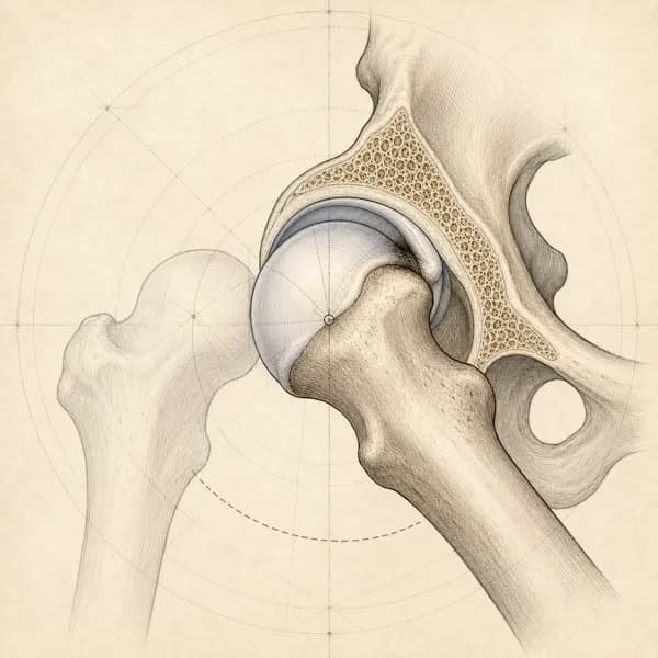
The joint: bone shape, labrum and cartilage
Wording about the ball and socket, the rim of cartilage around the socket, and the space between the bones.
A ball that is less round or a socket that covers more than usual on a hip X-ray or MRI, with the alpha angle and impingement wording that comes with it. How common the shapes are without pain, what they mean for arthritis, and what the trials show.
Wording this guide explains- Cam morphology, cam deformity, cam lesion
- Bump at the head-neck junction, aspherical femoral head, reduced head-neck offset, pistol grip deformity
- Alpha angle
- Pincer morphology, acetabular overcoverage, deep socket (coxa profunda, protrusio)
- Acetabular retroversion; crossover sign, posterior wall sign, ischial spine sign
- Lateral centre-edge angle (LCEA), centre-edge angle
- Mixed-type impingement, combined cam and pincer
- Femoroacetabular impingement (FAI), findings in keeping with FAI, FAI syndrome
- Herniation pit, fibrocystic change at the head-neck junction
- Os acetabulare, rim ossification, labral ossification
- Labral tear, labral degeneration, chondrolabral separation
- Joint space preserved or maintained; joint space narrowing; Tönnis grade
- Clinical correlation recommended
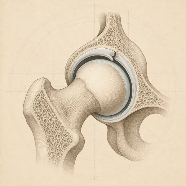
A frayed or split rim of the hip socket on an MRI or MR arthrogram. How often it appears in painless hips, which words on the report matter more, and what the trials say about physiotherapy and surgery.
Wording this guide explains- Labral tear, fraying, degeneration
- Anterosuperior
- Paralabral cyst
- Cam morphology, alpha angle
- Pincer morphology, over-coverage, retroversion
- Chondral or cartilage defect, delamination
- MR arthrogram
- Dysplasia, low centre-edge angle
- Gluteal tendinosis, trochanteric bursitis
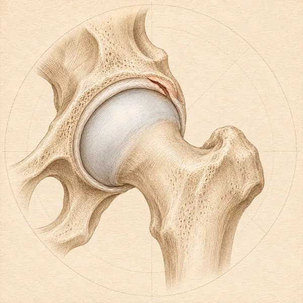
The gap between the ball and the socket looks thinner than expected on a hip X-ray. What superolateral, concentric and the severity words mean, how loosely it tracks pain, and what the guidelines and trials say.
Wording this guide explains- Superior or superolateral joint space narrowing
- Superomedial
- Medial or axial narrowing or migration
- Concentric, global, diffuse or uniform narrowing
- Minimal, mild or early
- Moderate
- Severe, marked, complete loss or “bone on bone”
- Loss of joint space, reduced joint space or joint space reduction
- Osteophytes or spurring
- Subchondral sclerosis
- Subchondral cysts or geodes
- Flattening, deformity or loss of roundness of the femoral head
- Acetabular protrusion or protrusio acetabuli
- Kellgren-Lawrence grade, Tönnis grade
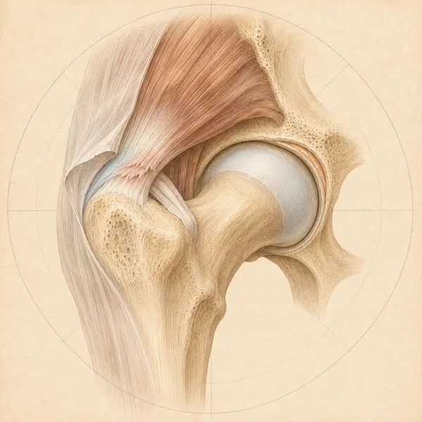
Tendons and bursae around the hip
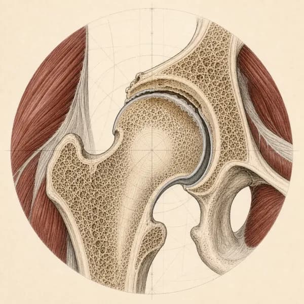
Wording about the tendons on the outside of the hip and at the sitting bone, and the fluid-filled sacs beside them.
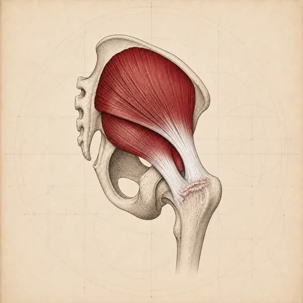
Thickened gluteal tendons, a gluteus medius tear or a thickened bursa on a hip ultrasound or MRI. Why the tendons are usually the source, how common these findings are without pain, and what the trials show.
Wording this guide explains- Gluteus medius, gluteus minimus, greater trochanter
- Tendinosis, tendinopathy, tendinitis
- Peritrochanteric edema or fluid, peritendinitis
- Trochanteric bursitis, bursal thickening, bursal fluid or distension
- Partial-thickness, interstitial or undersurface tear
- Full-thickness tear, retraction, avulsion
- Enthesopathy, cortical irregularity, enthesophyte
- Calcification, calcific tendinopathy
- Thickened iliotibial band
- Fatty atrophy or fatty infiltration
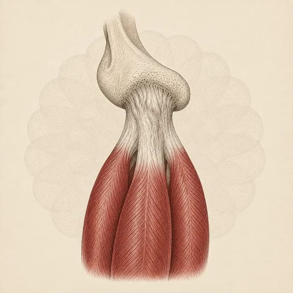
Thickened or changed hamstring tendons at the sitting bone on an MRI or ultrasound, with the partial-tear and bursa wording that comes with it. How common it is without symptoms and what the trials show.
Wording this guide explains- Hamstring origin, proximal hamstring, common hamstring tendon, ischial tuberosity
- Conjoint (conjoined) tendon; semimembranosus tendon
- Tendinosis, tendinopathy, tendonitis; mild, moderate, severe
- Thickening; increased signal or intrasubstance signal (MRI); hypoechoic area (ultrasound)
- Peritendinous edema or fluid, feathery edema
- Partial tear, partial-thickness tear, interstitial or intrasubstance tear; sickle sign
- Full-thickness or complete tear, avulsion, retraction
- Bone marrow edema at the ischial tuberosity
- Enthesopathy, cortical irregularity, spurring or enthesophyte
- Ischial bursitis, ischiogluteal bursitis
- Sciatic nerve close to, abutting or tethered by the tendon; perineural scarring
Related hip conditions
The condition pages behind the findings above: symptoms, treatment and recovery time.
Access, hours, and how to book
I see patients for hip pain at Endorphins Health & Wellness Centre in Burlington. The clinic serves people coming in from Burlington, Waterdown, Oakville, Hamilton, Flamborough, and Carlisle, with free parking on site and a ground-floor entrance.
- 4631 Palladium Way, Unit 6Burlington, ON L7M 0W9
- (905) 634-6000Direct insurance billing available. No physician referral needed.
Burlington hours
- Monday
- 1:30 to 7:00 PM
- Tuesday
- 3:30 to 8:00 PM
- Thursday
- 1:30 to 8:00 PM
- Wednesday
- 2:00 to 7:00 PM
- Friday
- 1:00 to 7:00 PM
- Tuesday
- 11:00 AM to 2:30 PM
- Saturday
- 12:00 to 3:00 PM