Cam and Pincer Morphology on a Hip Scan

Cam morphology on a hip X-ray or magnetic resonance imaging (MRI) report means the ball at the top of the thigh bone is less round than usual where it meets the neck of the bone. Pincer morphology means the socket covers more of the ball than usual. Reports also write femoroacetabular impingement (FAI), which is the idea that these shapes can pinch at the edge of the socket in deep bending. They describe the shape of the bones, not how much the hip hurts: in a review of 2,114 hips with no symptoms, 37 percent had cam morphology. The symptoms and the examination, read together with the picture, decide whether the shape explains your pain.

Assessing and treating hip pain in Burlington, Ontario.

When to see a doctor firstBroader Hip Pain Guide
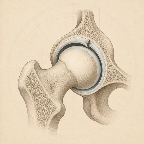
The ball and socket of the hip

What “cam” and “pincer” mean on a scan report

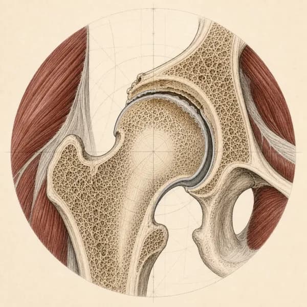
Two shapes, one on each side of the joint. The hip is a ball (the head of the thigh bone, or femur) in a socket (the acetabulum, part of the pelvis). Cam morphology is on the ball side: extra bone where the head meets the neck, so the head is not fully round. Pincer morphology is on the socket side: the rim reaches further over the ball than usual, all the way round or only at the front. A report that describes both calls it mixed.

Morphology, deformity, lesion. All three words describe the same shape. An international consensus panel agreed to use morphology, and to drop words with a negative ring such as lesion, abnormality and deformity (Dijkstra et al., 2022). Older reports and many studies still write cam deformity or cam lesion.

Impingement and the syndrome. Femoroacetabular impingement describes the ball and the rim of the socket meeting early in deep bending and turning. Femoroacetabular impingement syndrome is the diagnosis, and it needs three things together: symptoms that fit, signs on examination, and the shape on imaging (Griffin et al., 2016). A scan on its own cannot make it. The femoroacetabular impingement page covers the condition.

The words that come with it. These are the terms you are most likely to see with it on a hip or pelvis report.

On the reportWhat it means
Cam morphology, cam deformity, cam lesionExtra bone where the ball meets the neck of the thigh bone, so the ball is less round and the waist below it is shallower.
Bump at the head-neck junction, aspherical femoral head, reduced head-neck offset, pistol grip deformityOther ways of describing the cam shape: where the extra bone sits, and how it changes the outline of the ball and neck.
Alpha angleA measurement of how round the ball is where it meets the neck: the larger the angle, the less round. In the MRI study that introduced it, the angle averaged 74 degrees in 39 patients with signs of impingement and 42 degrees in 35 people without symptoms (Nötzli et al., 2002). Cut-offs differ between reports; the cohort studies on this page used more than 60 degrees to define cam morphology (Agricola et al., 2013).
Pincer morphology, acetabular overcoverage, deep socket (coxa profunda, protrusio)The socket covers more of the ball than usual. Studies have defined pincer morphology in many different ways, which is one reason its reported frequency varies so widely (Frank et al., 2015).
Acetabular retroversion; crossover sign, posterior wall sign, ischial spine signThe socket is angled to face slightly backward, so its front rim covers more of the ball. The three signs are features on a front-on pelvis X-ray that suggest it. They depend on how the pelvis was tilted when the picture was taken.
Lateral centre-edge angle (LCEA), centre-edge angleA measurement of how far the roof of the socket reaches over the ball. A large angle means more cover: a study of 18,935 hips used 40 degrees or more for moderate pincer morphology and 45 degrees or more for severe (Riedstra et al., 2026). A small angle means a shallow socket (hip dysplasia), which is a different problem; one trial used less than 20 degrees as its cut-off for dysplasia (Palmer et al., 2019).
Mixed-type impingement, combined cam and pincerBoth shapes are described in the same hip.
Femoroacetabular impingement (FAI), findings in keeping with FAI, FAI syndromeThe radiologist is saying the shape is one that can pinch. Whether it does in your hip depends on your symptoms and the examination (Griffin et al., 2016).
Herniation pit, fibrocystic change at the head-neck junctionA small, round, cyst-like area in the front of the neck of the thigh bone, sometimes noted alongside cam morphology.
Os acetabulare, rim ossification, labral ossificationA small separate piece of bone, or new bone, at the edge of the socket.
Labral tear, labral degeneration, chondrolabral separationChanges in the rim of cartilage (the labrum) around the socket. They are common in hips with and without pain; the labral tear on a hip MRI guide covers them.
Joint space preserved or maintained; joint space narrowing; Tönnis gradeHow much room there is between the ball and the socket, which reflects the cartilage. The Tönnis grade is a scale for arthritis on a hip X-ray, from 0 (none) to 3 (severe). The joint space narrowing in the hip guide covers this wording.
Clinical correlation recommendedThe radiologist is asking the person who examines you to decide whether the shape matches your symptoms. It is a standard line, not a sign that something was missed.

Cam morphology, cam deformity, cam lesion
Extra bone where the ball meets the neck of the thigh bone, so the ball is less round and the waist below it is shallower.
Bump at the head-neck junction, aspherical femoral head, reduced head-neck offset, pistol grip deformity
Other ways of describing the cam shape: where the extra bone sits, and how it changes the outline of the ball and neck.
Alpha angle
A measurement of how round the ball is where it meets the neck: the larger the angle, the less round. In the MRI study that introduced it, the angle averaged 74 degrees in 39 patients with signs of impingement and 42 degrees in 35 people without symptoms (Nötzli et al., 2002). Cut-offs differ between reports; the cohort studies on this page used more than 60 degrees to define cam morphology (Agricola et al., 2013).
Pincer morphology, acetabular overcoverage, deep socket (coxa profunda, protrusio)
The socket covers more of the ball than usual. Studies have defined pincer morphology in many different ways, which is one reason its reported frequency varies so widely (Frank et al., 2015).
Acetabular retroversion; crossover sign, posterior wall sign, ischial spine sign
The socket is angled to face slightly backward, so its front rim covers more of the ball. The three signs are features on a front-on pelvis X-ray that suggest it. They depend on how the pelvis was tilted when the picture was taken.
Lateral centre-edge angle (LCEA), centre-edge angle
A measurement of how far the roof of the socket reaches over the ball. A large angle means more cover: a study of 18,935 hips used 40 degrees or more for moderate pincer morphology and 45 degrees or more for severe (Riedstra et al., 2026). A small angle means a shallow socket (hip dysplasia), which is a different problem; one trial used less than 20 degrees as its cut-off for dysplasia (Palmer et al., 2019).
Mixed-type impingement, combined cam and pincer
Both shapes are described in the same hip.
Femoroacetabular impingement (FAI), findings in keeping with FAI, FAI syndrome
The radiologist is saying the shape is one that can pinch. Whether it does in your hip depends on your symptoms and the examination (Griffin et al., 2016).
Herniation pit, fibrocystic change at the head-neck junction
A small, round, cyst-like area in the front of the neck of the thigh bone, sometimes noted alongside cam morphology.
Os acetabulare, rim ossification, labral ossification
A small separate piece of bone, or new bone, at the edge of the socket.
Labral tear, labral degeneration, chondrolabral separation
Changes in the rim of cartilage (the labrum) around the socket. They are common in hips with and without pain; the labral tear on a hip MRI guide covers them.
Joint space preserved or maintained; joint space narrowing; Tönnis grade
How much room there is between the ball and the socket, which reflects the cartilage. The Tönnis grade is a scale for arthritis on a hip X-ray, from 0 (none) to 3 (severe). The joint space narrowing in the hip guide covers this wording.
Clinical correlation recommended
The radiologist is asking the person who examines you to decide whether the shape matches your symptoms. It is a standard line, not a sign that something was missed.

When to see a doctor first

Go to emergency

  • Groin or hip pain or a hot or swollen hip, together with fever, chills or feeling generally unwell

    Go to emergency now.

Get seen today

  • Sudden hip or groin pain with an inability to put weight on the leg, especially after a fall or if you are over 60

    Keep weight off the leg and get medical assessment today. A hip fracture needs to be ruled out.

  • A hot or swollen hip without fever, chills or feeling unwell

    Same-day medical assessment to rule out infection.

Check with your doctor

  • Hip pain in a child or teenager, with or without a limp or knee pain

    See your family doctor promptly. Hip problems in young people need a medical diagnosis before physiotherapy.

  • Deep hip pain with long-term steroid use, heavy alcohol use or sickle cell disease

    See your family doctor; these raise the chance of osteonecrosis of the femoral head.

  • Pain at night that does not ease, unexplained weight loss, or a history of cancer

    See your family doctor for a medical review before starting physiotherapy.

  • New numbness or weakness in the leg, or changes in bladder or bowel control

    Urgent medical assessment; this points to the spine or nerves rather than the hip joint.

What it does and does not tell you

These shapes are common in hips that do not hurt. A review pooled 26 studies of 2,114 hips with no symptoms, in people with an average age of 25. Cam morphology was reported in 37 percent, ranging from 7 to 100 percent between studies: about 55 percent in athletes and 23 percent in the general population. Pincer morphology was reported in 67 percent, with definitions that varied widely (Frank et al., 2015).

They are more marked, on average, in hips that do hurt. A review of 60 studies found cam morphology more often in people with symptoms and in athletes than in people without symptoms, and the average alpha angle was larger in the group with symptoms (Mascarenhas et al., 2016). The groups overlap, so a number on a report cannot sort one person into painful or not.

Cam morphology usually forms during growth. In 63 youth soccer players aged 12 to 19 who were X-rayed twice, about two and a half years apart, cam morphology developed gradually while the growth plate at the top of the thigh bone was still open, with no significant increase after it closed (Agricola et al., 2014). The consensus panel calls this primary cam morphology, to separate it from a cam shape left by an earlier hip condition (Dijkstra et al., 2022). In an adult the shape has usually been there since the teenage years, including through the years the hip did not hurt.

The scan does not make the diagnosis. An international group of 37 experts concluded that both the examination and imaging have limited ability to diagnose hip-related pain on their own, so the whole picture is needed. They recommend a front-on pelvis X-ray and a side view of the head and neck of the thigh bone as the first imaging, with MRI or other imaging only when more detail is needed, for example when planning surgery (Reiman et al., 2020).

It does not decide the treatment. What decides the plan is where the pain is, what brings it on and what the examination shows. Deep groin pain with sitting, squatting, getting out of a car or turning on the leg points to the joint. Pain on the outside of the hip or in the buttock usually points elsewhere, even with cam or pincer morphology on the report.

Cam and pincer morphology and arthritis later on

Cam morphology raises the odds of hip osteoarthritis. In 4,438 people aged 55 and over with no hip osteoarthritis at the start, followed for about nine years, those with cam morphology had roughly twice the odds of developing it on X-ray (Saberi Hosnijeh et al., 2017). In a Dutch cohort of 1,002 people aged 45 to 65 with early symptoms of osteoarthritis, an alpha angle above 60 degrees went with 3.7 times the odds of end-stage osteoarthritis within five years, and an angle above 83 degrees with 9.7 times the odds (Agricola et al., 2013).

Most hips with cam morphology still did not get there. In that Dutch cohort, 2.76 percent of hips reached end-stage osteoarthritis within five years. The hips most at risk had a severe cam shape together with reduced inward rotation on examination: about half of those reached end-stage osteoarthritis (Agricola et al., 2013). Ten years on, hips that began with pain, limited rotation and cam morphology together had much higher odds of osteoarthritis than hips with none of the three, but only 21 of 1,386 hips started in that group (Agricola et al., 2024).

For pincer morphology the link is weaker. Pincer morphology did not raise the risk of osteoarthritis in the study of people aged 55 and over (Saberi Hosnijeh et al., 2017). In 18,935 hips followed for up to eight years, moderate pincer morphology was not linked with new osteoarthritis, and severe pincer morphology went with 1.5 times the odds; 1.9 percent of all the hips developed it (Riedstra et al., 2026).

What that means for one hip. These studies describe groups. They show that the shape is one risk factor among several, and that most hips with it did not develop severe arthritis in the years they were followed. Whether reshaping the bone lowers that risk has not been shown. In a trial that measured cartilage health on MRI a year after surgery or physiotherapist-led care, the two groups did not differ significantly (Hunter et al., 2021).

Why deep bending, sitting and twisting bring it on

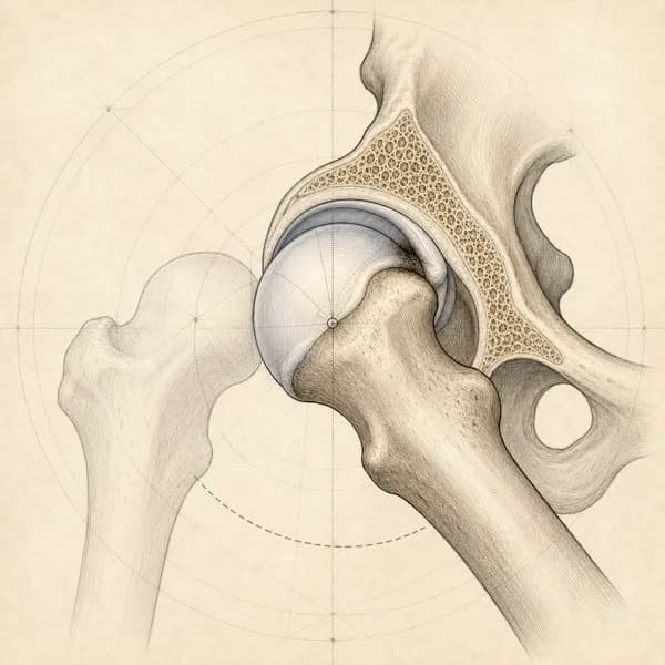
As the hip bends deeply and turns inward, the front of the neck of the thigh bone moves toward the front rim of the socket. With extra bone at the head and neck junction, or a rim that reaches further over the ball, the two can meet earlier in the movement (Nötzli et al., 2002). The labrum and the cartilage at the edge of the socket sit between them, which is why reports that mention cam or pincer morphology often mention the labrum as well.

That matches the usual triggers: low chairs and long drives, deep squats, getting in and out of a car, and cutting or pivoting in sport. The shape is the same on good days and bad days, so how much the hip is asked to do in those positions, and how irritable the joint is at the time, account for much of the variation. Sitting higher, breaking up long periods of sitting and keeping squats above the depth that pinches may ease symptoms while strength is built up.

What the evidence says about treatment

The consensus statement lists three suitable treatments for femoroacetabular impingement syndrome: conservative care, rehabilitation, and arthroscopic or open surgery (Griffin et al., 2016). The trials below compare rehabilitation with arthroscopy. Morphology found on a scan of a hip that does not hurt is not a reason for either.

  • Exercise-based rehabilitation. An international consensus recommends exercise-based treatment for hip-related pain, for at least three months, and recommends staying physically active, sport included. The same group notes that strong evidence for physiotherapist-led treatment is lacking and that the best type and amount of exercise are not established (Kemp et al., 2020).
  • Arthroscopy compared with physiotherapist-led care, UK FASHIoN. In 348 adults, both groups improved over 12 months. On a 100-point scale of hip-related quality of life, scores rose from about 39 to 59 with arthroscopy and from about 36 to 50 with a personalised physiotherapy program. After adjustment, arthroscopy scored 6.8 points higher, just above the smallest difference the trial counted as important, which was 6.1 points (Griffin et al., 2018).
  • Arthroscopy compared with physiotherapy, FAIT. In 222 adults aged 18 to 60, scores for everyday activities eight months after joining were 10 points higher, on a 100-point scale, with arthroscopy than with a physiotherapy program of up to eight sessions (Palmer et al., 2019).
  • A trial with a different result. In 80 military patients there was no significant difference between surgery and physiotherapy at two years. Of the 40 assigned to physiotherapy, 28 went on to have surgery, and analysing by the treatment people received did not change the result (Mansell et al., 2018).
  • Taken together. A pooled analysis of three trials found a benefit for arthroscopy over non-surgical care in quality of life and everyday activities, and no significant difference in sports function (Mok et al., 2021). People improved in both arms of these trials, the differences were modest on average, and none of the trials compared surgery with a sham operation. The choice is a shared one, made with a surgeon when surgery is being considered.
  • How long to give rehabilitation. The consensus recommendation is at least three months of exercise-based treatment (Kemp et al., 2020). Trying rehabilitation first did not close off surgery in the trials: in the military trial most of the physiotherapy group later chose it (Mansell et al., 2018).

How I assess and treat a hip with this on the report

I start with where the pain is and what brings it on: deep groin pain with sitting, squatting, getting out of a car or turning on the leg points to the joint, while pain on the outside of the hip or in the buttock usually points elsewhere. I ask about sport and training through the teenage years, how long it has been going on and what it stops you doing.

On examination I measure hip range of motion, inward rotation in particular, and test the movements that pinch at the front of the hip. A test that bends the hip, brings it across the body and turns it inward is useful mainly when it does not hurt, which makes the joint a less likely source (Reiman et al., 2020). I test strength in the hip and trunk and screen the lower back and the sacroiliac joint, because they refer pain to the same area. Then I read the report alongside what I found.

The plan is usually strength for the hip and trunk, work on the movements that provoke the hip so you can do them without pinching, and changes to the loads that flare it, built up toward the sport or activity you want back. Hands-on treatment can help with pain and movement alongside that. The femoroacetabular impingement page sets out the stages in detail, and the labral tear or impingement comparison covers how the two overlap.

If the hip is not responding after a fair course, usually about three months, the next step is a surgical opinion through your family doctor or a sports medicine physician. I cannot order imaging; when imaging would change the plan, I flag it to your family doctor or a sports medicine physician and refer you.

Cam and pincer questions I hear most

What does cam morphology mean on a hip X-ray?

The ball at the top of the thigh bone is less round than usual where it meets the neck of the bone. It describes a shape, not an injury, and it is common in hips that do not hurt: a review of 2,114 hips with no symptoms found it in 37 percent. Whether it matters depends on your symptoms and the examination.

What does pincer morphology mean?

The socket covers more of the ball than usual, either all the way round or only at the front. Reports may call it overcoverage or acetabular retroversion. Studies define it in many different ways, and it is frequently reported in hips with no symptoms.

Is cam or pincer morphology the same as femoroacetabular impingement?

Not quite. Cam and pincer are the shapes. Femoroacetabular impingement syndrome is the diagnosis, and it needs fitting symptoms, signs on examination and the shape on imaging together. Many people have the shape without the syndrome.

What is a normal alpha angle?

There is no single agreed cut-off. The alpha angle measures how round the ball is where it meets the neck, and a larger angle means less round. In the study that introduced it, the angle averaged 42 degrees in people without symptoms and 74 degrees in patients with signs of impingement. Several large studies used more than 60 degrees to define cam morphology. Reports measure it on different views, so the same hip can give different numbers.

Will cam morphology give me arthritis?

It raises the odds, but most hips with it did not develop severe arthritis in the studies that followed people over time. In one Dutch cohort, under 3 percent of hips reached end-stage osteoarthritis within five years. The hips most at risk had a severe cam shape together with reduced inward rotation on examination.

Does cam or pincer morphology need surgery?

Not on its own. Surgery is considered for femoroacetabular impingement syndrome, usually after a course of rehabilitation has not helped enough. In the trials, people improved with both arthroscopy and physiotherapy, with somewhat better scores on average after arthroscopy in two trials and no significant difference in a third. The decision is made with a surgeon.

Can physiotherapy change the shape of the bone?

No. Exercise does not change the shape. The aim is a hip that is stronger, moves well within the range it has and copes with the loads you need, so that the shape matters less. An international consensus recommends at least three months of exercise-based treatment for hip-related pain.

Should I stop sport or squatting if my report says cam morphology?

Usually not. The consensus recommendation for hip-related pain is to stay physically active, sport included. If deep squats or long periods of sitting bring on the pain, adjusting depth, load or position for a while is often enough while strength is built up, and the adjustments are eased as the hip settles.

Do I need an MRI if my X-ray shows cam or pincer morphology?

Not always. Consensus recommendations name a front-on pelvis X-ray and a side view of the head and neck of the thigh bone as the first imaging, with MRI when more detail of the bone or soft tissue is needed, for example when surgery is being planned. I cannot order imaging; when imaging would change the plan, I flag it to your family doctor or a sports medicine physician and refer you.

Evidence this page is built on

The main sources behind this guide. Other studies are named in the text where they are used.

2015 Frank JM et al., Arthroscopy

Prevalence of femoroacetabular impingement imaging findings in asymptomatic volunteers: a systematic review

26 studies, 2,114 hips with no symptoms, average age 25. Cam morphology in 37 percent (7 to 100 percent between studies): 54.8 percent in athletes and 23.1 percent in the general population. Pincer morphology in 67 percent, poorly and variably defined. The average alpha angle was 54 degrees.

2016 Mascarenhas VV et al., European Journal of Radiology

Imaging prevalence of femoroacetabular impingement in symptomatic patients, athletes, and asymptomatic individuals: a systematic review

60 studies. Cam morphology was more common in athletes and in people with symptoms than in people without symptoms, and the average alpha angle was larger in the group with symptoms. Imaging signs were common in all three groups.

2002 Nötzli HP et al., Journal of Bone and Joint Surgery (British)

The contour of the femoral head-neck junction as a predictor for the risk of anterior impingement

The study that introduced the alpha angle. On MRI it averaged 74 degrees in 39 patients with groin pain, reduced inward rotation and a positive impingement test, and 42 degrees in 35 people without symptoms.

2016 Griffin DR et al., British Journal of Sports Medicine

The Warwick Agreement on femoroacetabular impingement syndrome: an international consensus statement

Consensus of 22 panel members and a patient from 9 countries. The syndrome is diagnosed from symptoms, clinical signs and imaging findings together. Suitable treatments are conservative care, rehabilitation, and arthroscopic or open surgery.

Show all 17 sources

2022 Dijkstra HP et al., British Journal of Sports Medicine

Oxford consensus on primary cam morphology and femoroacetabular impingement syndrome: part 1, definitions, terminology, taxonomy and imaging outcomes

A panel of 65 from 18 countries agreed a definition of primary cam morphology, agreed to use “morphology” in place of “lesion”, “abnormality” or “deformity”, and separated primary from secondary cam morphology.

2020 Reiman MP et al., British Journal of Sports Medicine

Consensus recommendations on the classification, definition and diagnostic criteria of hip-related pain in young and middle-aged active adults

Panel of 37 experts. Examination and imaging each have limited diagnostic value alone. A front-on pelvis X-ray and a side view of the femoral head and neck are the first imaging of choice; advanced imaging only when more detail is needed. A negative flexion, adduction and internal rotation test helps rule out hip-related pain.

2014 Agricola R et al., American Journal of Sports Medicine

A cam deformity is gradually acquired during skeletal maturation in adolescent and young male soccer players: a prospective study with minimum 2-year follow-up

63 youth soccer players aged 12 to 19, X-rayed at the start and about 2.4 years later. Cam morphology increased while the growth plate was open, with no significant increase after it closed.

2013 Agricola R et al., Annals of the Rheumatic Diseases

Cam impingement causes osteoarthritis of the hip: a nationwide prospective cohort study (CHECK)

1,002 people with early symptoms, X-rayed over five years. 2.76 percent of hips reached end-stage osteoarthritis. An alpha angle above 60 degrees gave an adjusted odds ratio of 3.67 and above 83 degrees 9.66. Severe cam morphology with reduced inward rotation had a positive predictive value of 52.6 percent.

2024 Agricola R et al., British Journal of Sports Medicine

Femoroacetabular impingement syndrome in middle-aged individuals is strongly associated with the development of hip osteoarthritis within 10-year follow-up

1,386 hips in people aged 45 to 65. The 21 hips with pain, limited inward rotation and cam morphology together had an adjusted odds ratio of 6.85 for osteoarthritis on X-ray within 10 years, compared with hips that had none of the three.

2017 Saberi Hosnijeh F et al., Arthritis & Rheumatology

Cam deformity and acetabular dysplasia as risk factors for hip osteoarthritis

4,438 people aged 55 and over without hip osteoarthritis, followed for an average of 9.2 years. Cam morphology and a shallow socket each roughly doubled the odds of osteoarthritis; pincer morphology did not raise it. The links were driven by the younger participants.

2026 Riedstra N et al., British Journal of Sports Medicine

Severe pincer morphology is associated with incident hip osteoarthritis: prospective individual participant data from 18 935 hips from the World COACH consortium

18,935 hips from nine cohorts, followed for four to eight years; 1.9 percent developed osteoarthritis on X-ray. Moderate pincer morphology (lateral centre-edge angle of 40 degrees or more) was not linked with it; severe (45 degrees or more) had an odds ratio of 1.50.

2018 Griffin DR et al., The Lancet

Hip arthroscopy versus best conservative care for the treatment of femoroacetabular impingement syndrome (UK FASHIoN)

348 adults at 23 hospitals. At 12 months, quality of life scores had risen from 39.2 to 58.8 with arthroscopy and from 35.6 to 49.7 with personalised hip therapy; the adjusted difference was 6.8 points in favour of arthroscopy, against a minimum important difference of 6.1.

2019 Palmer AJR et al., BMJ

Arthroscopic hip surgery compared with physiotherapy and activity modification for the treatment of symptomatic femoroacetabular impingement (FAIT)

222 adults aged 18 to 60 at seven centres. Eight months after randomisation, the score for activities of daily living was 10.0 points higher with arthroscopy than with a physiotherapy program of up to eight sessions. No serious adverse events in either group.

2018 Mansell NS et al., American Journal of Sports Medicine

Arthroscopic surgery or physical therapy for patients with femoroacetabular impingement syndrome: a randomized controlled trial with 2-year follow-up

80 military patients. No significant difference between surgery and physiotherapy at two years; 28 of 40 assigned to rehabilitation crossed over to surgery without changing the result.

2021 Hunter DJ et al., BMC Musculoskeletal Disorders

Multi-centre randomised controlled trial comparing arthroscopic hip surgery to physiotherapist-led care for femoroacetabular impingement syndrome on hip cartilage metabolism (Australian FASHIoN)

99 participants, average age 33. The MRI measure of cartilage health at 12 months did not differ significantly between the groups. Quality of life improved in both, by 14 points more with arthroscopy.

2021 Mok TN et al., Orthopaedic Surgery

Arthroscopic hip surgery versus conservative therapy on femoroacetabular impingement syndrome: a meta-analysis of RCTs

Three randomised trials. Arthroscopy gave more improvement than non-surgical care in quality of life (575 participants) and activities of daily living (262 participants); the difference in sports function was not significant.

2020 Kemp JL et al., British Journal of Sports Medicine

Physiotherapist-led treatment for young to middle-aged active adults with hip-related pain: consensus recommendations

Consensus of 38 researchers and clinicians. Exercise-based treatment is recommended, for at least three months, along with physical activity, shared decision-making and education. The accompanying review found strong evidence for physiotherapist-led treatment lacking.

Access, hours, and how to book

I see patients for hip pain at Endorphins Health & Wellness Centre in Burlington. The clinic serves people coming in from Burlington, Waterdown, Oakville, Hamilton, Flamborough, and Carlisle, with free parking on site and a ground-floor entrance.

  • 4631 Palladium Way, Unit 6Burlington, ON L7M 0W9
  • (905) 634-6000Direct insurance billing available. No physician referral needed.

Burlington hours

Endorphins Health & Wellness Centre
4631 Palladium Way
Monday
1:30 to 7:00 PM
Tuesday
3:30 to 8:00 PM
Thursday
1:30 to 8:00 PM
Headon Physio
1387 Walkers Line
Wednesday
2:00 to 7:00 PM
Friday
1:00 to 7:00 PM
PhysioMax Wellness
1035 Brant Street
Tuesday
11:00 AM to 2:30 PM
Saturday
12:00 to 3:00 PM