What Your Lumbar Spine MRI Report Means

A lumbar MRI report describes each level of the lower back in turn, in language written for the doctor who ordered it. Most of its phrases describe how the discs, joints and nerve openings look. Many of those findings are common in people with no back pain at all, so the report has to be read alongside your symptoms and an exam.

Assessing and treating lower back pain in Burlington, Ontario.

When to see a doctor firstBroader Low Back Pain Guide
The lower back

How a lumbar report is laid out

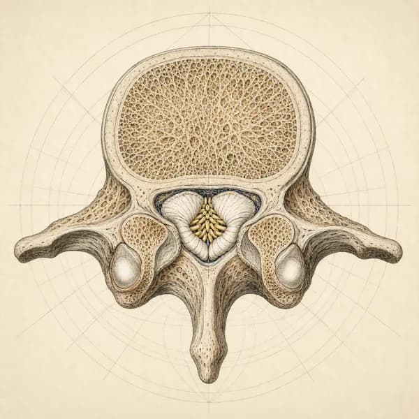
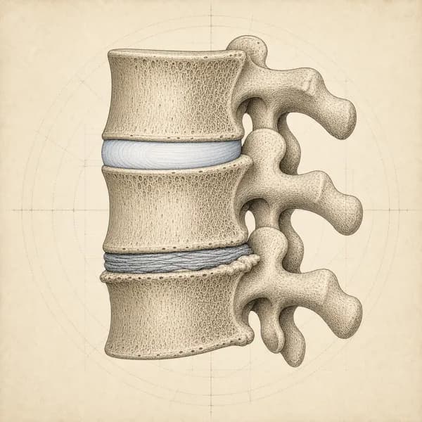
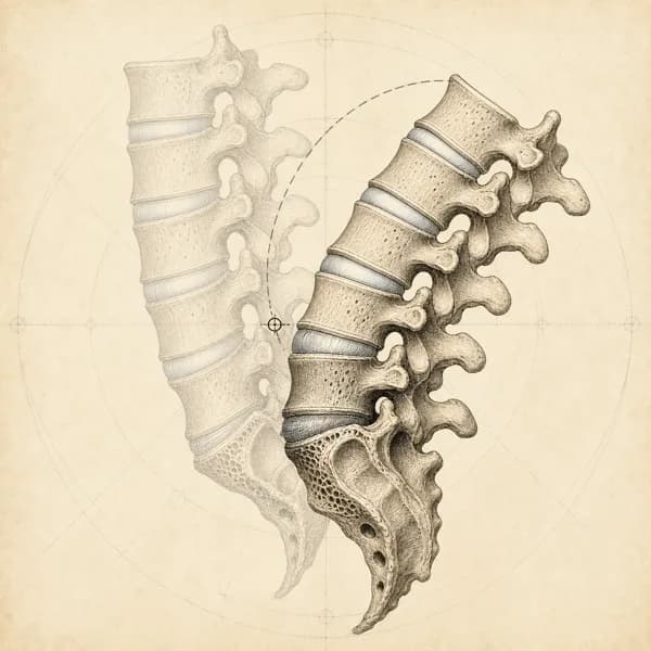
The lower back has five lumbar vertebrae, L1 to L5, sitting on the sacrum (S1). A disc is named by the two bones it sits between, so "L4-L5" is the disc between the fourth and fifth lumbar vertebrae. Most reports work down the spine one level at a time and end with an "Impression", which is the radiologist's short summary.

Words such as mild, moderate and severe describe how a finding looks on the picture. They are not a pain score, and they do not say how the back works.

When to see a doctor first

Go to emergency

  • New difficulty passing urine or controlling your bladder or bowels, or numbness around the groin, genitals or inner thighs

    Go to emergency now. These can be signs of pressure on the nerves at the base of the spine (cauda equina syndrome).

Get seen today

  • Weakness in a leg or foot that is new or getting worse, or a foot that drags

    Seek same-day medical assessment.

  • Back pain with fever, chills or feeling generally unwell

    Seek same-day medical assessment to rule out infection.

Check with your doctor

  • Back pain after a significant fall or accident, or after a minor fall if you have osteoporosis or take long-term steroid medication

    Seek medical assessment to rule out a fracture before starting rehab.

  • A history of cancer with new back pain, or back pain with unexplained weight loss

    See your family doctor promptly.

Words about the discs

On the reportWhat it means
Disc desiccationThe disc holds less water than it once did and looks darker on the scan. A common age-related change.
Loss of disc heightThe space the disc occupies between the two bones is shorter than expected. Often reported together with desiccation as "degenerative disc disease".
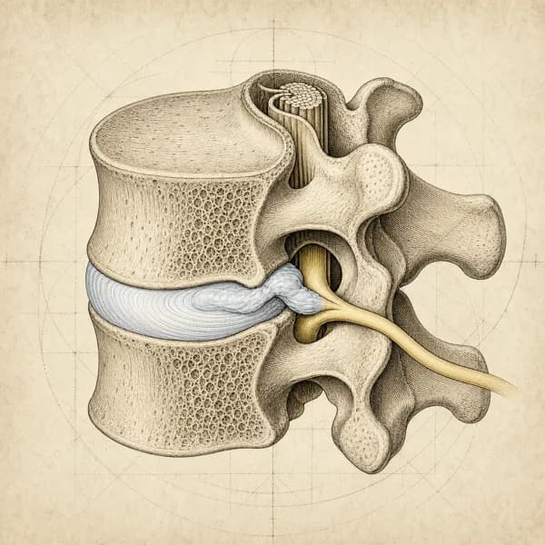
Disc bulgeThe outer edge of the disc extends a little past the bone, around a wide part of its rim. See the guide to disc bulge, protrusion and extrusion.
Protrusion, extrusionTwo shapes of disc herniation, where disc material is displaced at one spot. The same guide covers how they differ.
Annular fissure or annular tearA small split in the outer ring of the disc. Common on scans of people without pain.
Endplate (Modic) changesA change in the bone next to a disc, reported as type 1, 2 or 3 according to how it looks.
Schmorl's nodeA small dent where disc material has pressed into the bone above or below. Usually an incidental finding.
Disc desiccation
The disc holds less water than it once did and looks darker on the scan. A common age-related change.
Loss of disc height
The space the disc occupies between the two bones is shorter than expected. Often reported together with desiccation as "degenerative disc disease".
Disc bulge
The outer edge of the disc extends a little past the bone, around a wide part of its rim. See the guide to disc bulge, protrusion and extrusion.
Protrusion, extrusion
Two shapes of disc herniation, where disc material is displaced at one spot. The same guide covers how they differ.
Annular fissure or annular tear
A small split in the outer ring of the disc. Common on scans of people without pain.
Endplate (Modic) changes
A change in the bone next to a disc, reported as type 1, 2 or 3 according to how it looks.
Schmorl's node
A small dent where disc material has pressed into the bone above or below. Usually an incidental finding.

Words about the joints and nerve openings

On the reportWhat it means
Facet arthropathy or facet hypertrophyWear-related change, or enlargement, of the small joints at the back of the spine.
Ligamentum flavum thickeningA ligament inside the spinal canal looks thicker than usual. It can contribute to narrowing of the canal.
Central canal stenosisThe main channel that the nerves run through is narrower than expected. More on the spinal stenosis page.
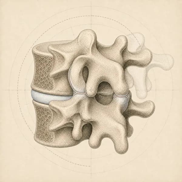
Foraminal narrowing or foraminal stenosisThe side opening where a nerve root leaves the spine is narrower than expected.
Lateral recess narrowingNarrowing in the corner of the canal, where a nerve root travels just before it exits.
Contacting, abutting or compressing a nerve rootContact or abutment means tissue touches the nerve root. Compression means it presses on the nerve and may change its shape. Your symptoms and the exam help assess whether the finding is contributing to the problem.
SpondylolisthesisOne vertebra sits forward or back on the one below. It is graded by how far it has moved.
SpondylosisA general word for age-related change in the spine.
Facet arthropathy or facet hypertrophy
Wear-related change, or enlargement, of the small joints at the back of the spine.
Ligamentum flavum thickening
A ligament inside the spinal canal looks thicker than usual. It can contribute to narrowing of the canal.
Central canal stenosis
The main channel that the nerves run through is narrower than expected. More on the spinal stenosis page.
Foraminal narrowing or foraminal stenosis
The side opening where a nerve root leaves the spine is narrower than expected.
Lateral recess narrowing
Narrowing in the corner of the canal, where a nerve root travels just before it exits.
Contacting, abutting or compressing a nerve root
Contact or abutment means tissue touches the nerve root. Compression means it presses on the nerve and may change its shape. Your symptoms and the exam help assess whether the finding is contributing to the problem.
Spondylolisthesis
One vertebra sits forward or back on the one below. It is graded by how far it has moved.
Spondylosis
A general word for age-related change in the spine.

What the report does and does not tell you

Many findings are common without pain. A review of 33 studies, covering 3,110 people with no back pain, estimated that disc degeneration shows on scans in 37% of 20-year-olds and 96% of 80-year-olds. Its estimates for disc bulges were 30% of 20-year-olds and 84% of 80-year-olds, and for disc protrusions 29% and 43%.

So a finding is not automatically the cause. A report can describe several changes at several levels. Comparing the report with your symptoms and the exam helps judge which of them may be contributing. This is also why guidelines advise against routine imaging for low back pain without signs of a serious or specific condition.

How I use a report in clinic

I compare the report with your history and the exam, including where the pain travels, which movements change it, and your strength, reflexes and sensation. This helps judge which findings may be contributing to your symptoms and whether further assessment is needed.

I cannot order imaging. When a scan, or a repeat scan, would change the plan, I flag it to your family doctor or specialist and refer you.

Questions about lumbar MRI reports

Does my MRI explain my back pain?

Sometimes, and often only in part. Many findings on a lumbar MRI also occur in people without pain. Comparing the scan with your symptoms and the exam helps assess which findings may be contributing.

Is degenerative disc disease a disease?

Despite the name, it is a description of how the discs look: less water, less height. In a review of people with no back pain, disc degeneration was estimated to be present in 37% of 20-year-olds and 96% of 80-year-olds.

What do mild, moderate and severe mean on the report?

They describe how marked a finding looks on the scan. They do not grade pain, and a finding called severe on the picture can cause little trouble, while a mild one can be the source of symptoms.

My report lists several levels. Is that worse?

Not necessarily. Age-related changes are often seen at more than one level. What matters is whether any of them fits your symptoms.

Do I need surgery?

An MRI finding alone does not decide that. Most back and leg pain is managed without surgery. A surgical opinion may be considered when leg pain keeps causing substantial pain or limiting what you can do despite non-surgical care, and the scan findings fit the symptoms. New or worsening leg weakness needs urgent medical assessment. New difficulty passing urine, loss of bladder or bowel control, or numbness around the genitals or anus needs emergency assessment.

Is it safe to exercise with these findings?

For most people, yes, and staying active is part of usual care for low back pain. I set the starting point and the progression from the exam and from how your back responds.

Evidence this page is built on

The main sources behind this guide. Other studies are named in the text where they are used.

2015 Brinjikji W, Luetmer PH, Comstock B, et al. American Journal of Neuroradiology

Systematic literature review of imaging features of spinal degeneration in asymptomatic populations

In 33 studies of 3,110 people without symptoms, disc degeneration was present in 37% of 20-year-olds and 96% of 80-year-olds, disc bulge in 30% and 84%, disc protrusion in 29% and 43%, and annular fissure in 19% and 29%.
2014 Fardon DF, Williams AL, Dohring EJ, et al. The Spine Journal

Lumbar disc nomenclature: version 2.0

The consensus recommendations of three North American spine and radiology societies on the standard terms for normal and abnormal lumbar discs, including bulge, protrusion, extrusion and sequestration.

2011 Chou R, Qaseem A, Owens DK, Shekelle P. Annals of Internal Medicine

Diagnostic imaging for low back pain: advice for high-value health care from the American College of Physicians

Advises imaging for low back pain only with severe progressive neurologic deficits or signs of a serious or specific underlying condition, and reports that routine imaging in other patients is not associated with meaningful benefit and can lead to harms.

Access, hours, and how to book

I see patients for lower back pain at Endorphins Health & Wellness Centre in Burlington. The clinic serves people coming in from Burlington, Waterdown, Oakville, Hamilton, Flamborough, and Carlisle, with free parking on site and a ground-floor entrance.

  • 4631 Palladium Way, Unit 6Burlington, ON L7M 0W9
  • (905) 634-6000Direct insurance billing available. No physician referral needed.

Burlington hours

Endorphins Health & Wellness Centre
4631 Palladium Way
Monday
1:30 to 7:00 PM
Tuesday
3:30 to 8:00 PM
Thursday
1:30 to 8:00 PM
Headon Physio
1387 Walkers Line
Wednesday
2:00 to 7:00 PM
Friday
1:00 to 7:00 PM
PhysioMax Wellness
1035 Brant Street
Tuesday
11:00 AM to 2:30 PM
Saturday
12:00 to 3:00 PM