
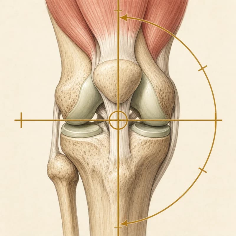
What Your Knee MRI Report Means
A knee MRI report lists what the scan shows, one structure at a time, in radiology shorthand. This page gathers the phrases that appear most often, gives each a short plain-language meaning, and links to its full guide. A report describes the picture. It does not say how much the knee hurts or what it can still do, and several of these findings are common in knees with no pain.
Assessing and treating knee pain at the Burlington clinic. Convenient for Waterdown, Oakville, Hamilton, Flamborough, and Carlisle residents.
When to see a doctor firstBroader Knee Pain GuideHow a knee MRI report is laid out
Findings and impression. Most reports have two parts. The findings section goes through the knee one structure at a time: the menisci, the ligaments, the cartilage, the bone, the tendons and the fluid in the joint. The impression at the end is the radiologist’s short summary of what stood out.
Size and grade words. Words such as trace, mild, moderate and severe, and grades such as 1 to 4, are the radiologist’s estimate of how much is there. They describe the picture. They are not a measure of pain, and radiologists do not all draw the lines in the same place.
What the report cannot tell you. A report cannot say which finding, if any, is the source of the pain. That takes the history of the knee and an examination, read together with the scan. The guides below give, for each phrase, what it means, how often it shows up in knees that do not hurt, and when it needs a doctor first.
When to see a doctor first
Go to emergency
After an injury, the kneecap is still out of place, the knee looks deformed or dislocated, or the foot is cold, pale or numb
Go to emergency now. Do not try to put the kneecap or knee back yourself, and do not walk on the leg.
A hot, red or swollen knee with fever, chills or feeling generally unwell
Go to emergency now. This can be a joint infection.
A limping child or teenager with sudden hip, thigh or knee pain, who cannot put weight on the leg, whose leg looks deformed, or who has a fever and feels unwell
Go to emergency now.
A suspected slipped growth plate at the hip (slipped capital femoral epiphysis, or SCFE)
Do not let the child walk on the leg, and seek emergency assessment today. In a teenager this can show up as a limp with hip, groin, thigh or knee pain, even when they can still walk.
Sudden shortness of breath or chest pain, coughing up blood or feeling faint, especially with a painful or swollen calf
Go to emergency now, or call 911. A clot that has travelled to the lungs (pulmonary embolism) needs to be ruled out.
Get seen today
Unable to bear weight after trauma, or the knee gave way with a pop and immediate swelling
Get same-day assessment at urgent care or emergency for a significant knee injury. The clinician examines the knee and, where appropriate, uses the Ottawa Knee Rules to decide whether an X-ray is needed for a fracture. Ligament or cartilage injuries need their own examination and sometimes later imaging.
A newly hot, red or swollen knee without fever, chills or feeling unwell
Get medical assessment the same day to rule out a joint infection or gout. Infection is possible even without a fever.
Locked knee that cannot be straightened or bent fully
A mechanically locked knee needs orthopaedic review before rehabilitation can progress safely. Get it assessed the same day by your family doctor, urgent care or emergency; it needs an urgent orthopaedic referral.
Calf pain, warmth, or swelling behind the knee, particularly after travel or surgery
Seek same-day medical assessment to rule out a blood clot (deep vein thrombosis) before starting physiotherapy.
Numbness, weakness, or foot drop alongside the knee pain that is getting worse
Get medical assessment the same day to check for a nerve injury or a nerve root problem in the lower back.
Check with your doctor
A child or teenager with a new limp that has no clear explanation
Get a prompt medical assessment before starting exercises. A hip problem can be felt only in the thigh or knee.
Unexplained weight loss, night pain, or a history of cancer with new knee pain
See your family physician for medical workup before starting physiotherapy.
Fluid and cysts
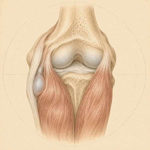
Wording about extra fluid in the joint and the pocket it can fill behind the knee.
Extra fluid inside the knee joint, seen in the pouch above the kneecap. What the size words on a report mean, the usual causes by how quickly the swelling came on, and when to see a doctor first.
Wording this guide explains- Suprapatellar effusion, joint effusion
- Trace or minimal
- Small or mild
- Moderate
- Large or marked
- Suprapatellar pouch, recess or bursa
A pocket of fluid behind the knee on an ultrasound or MRI, usually fed by a problem inside the joint. What the report wording means, why new calf swelling needs a same-day check, and how it is treated.
Wording this guide explains- Baker's cyst, popliteal cyst
- Distended semimembranosus-gastrocnemius bursa
- Communicating with the joint, neck
- Simple or anechoic
- Complex, septations, debris, loose bodies
- Ruptured, leaking or dissecting
- Joint effusion
- Tear of the posterior horn of the medial meniscus
- Osteoarthritis, cartilage loss, osteophytes
- Size in mm or cm
Cartilage and bone
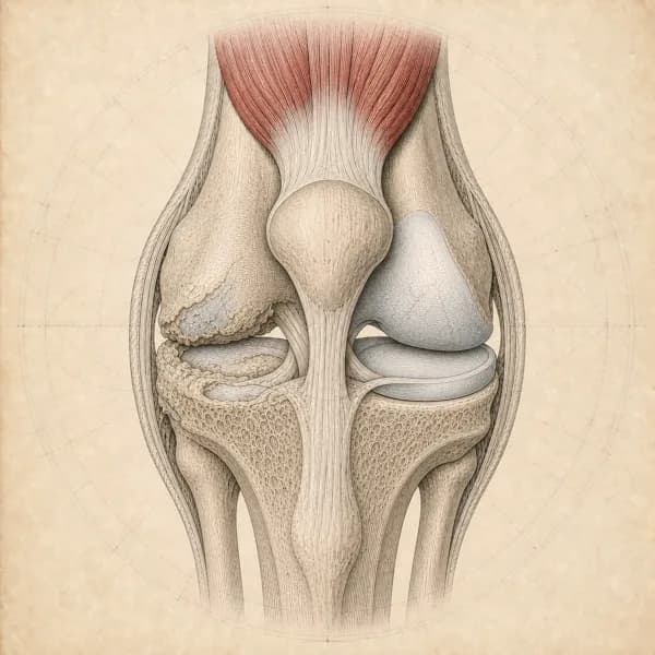
Wording about the joint surfaces and the bone under them, on an MRI or an X-ray.
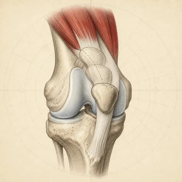
Softened or worn cartilage behind the kneecap on an MRI. What the grades describe, why people with and without kneecap pain show the same findings, and what the guidelines recommend.
Wording this guide explains- Grade 1
- Grade 2
- Grade 3
- Grade 4
- Medial or lateral facet, apex, trochlea
- Focal defect, fissure, delamination
- Subchondral bone marrow edema
- Patellar tilt, lateral subluxation, trochlear dysplasia
- Hoffa fat pad edema
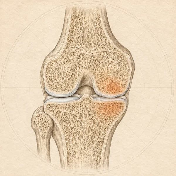
A bright patch of bone under the joint surface on an MRI, also called a bone marrow lesion. Why it is common in painless knees, what it means next to arthritis or an injury, and the signs that need a doctor first.
Wording this guide explains- Bone marrow lesion (BML)
- Subchondral
- Medial or lateral femoral condyle, tibial plateau, patella
- Focal or diffuse, mild, moderate or extensive
- Bone contusion or bone bruise
- Stress reaction or stress response
- Subchondral insufficiency fracture
- Osteonecrosis or SONK
- With overlying cartilage loss or chondral defect
- Transient or migratory
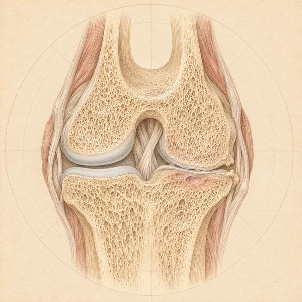
A small pocket in the bone under the joint surface on a knee X-ray or MRI, also written as cystic change or a geode. How it relates to arthritis and pain, and the report words that need a doctor first.
Wording this guide explains- Subchondral cyst, subchondral bone cyst
- Subchondral cystic change, cyst-like lesion, cystlike
- Geode
- Small, tiny, few, multiple
- Medial or lateral femoral condyle, tibial plateau, patella, trochlea
- Subchondral sclerosis
- Joint space narrowing, osteophytes
- Bone marrow edema or bone marrow lesion
- Well-defined, degenerative, in keeping with osteoarthritis
- Expansile, aggressive, atypical, further imaging recommended
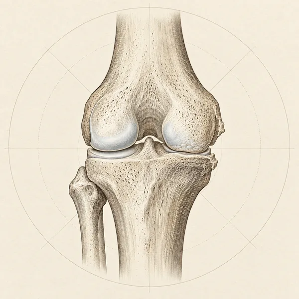
The gap between the bones looks thinner than expected on an X-ray. What medial, tricompartmental and the severity words mean, how it relates to pain, and what the guidelines recommend.
Wording this guide explains- Tibiofemoral joint space narrowing
- Medial compartment
- Lateral compartment
- Patellofemoral
- Bicompartmental or tricompartmental
- Joint space reduction, loss of joint space or reduced joint space
- Minimal, mild or early
- Moderate
- Severe, marked or “bone on bone”
- A percentage, such as “less than 50% joint space narrowing”
- Osteophytes or spurring
- Subchondral sclerosis
- Joint effusion
- Standing or weight-bearing view
Meniscus
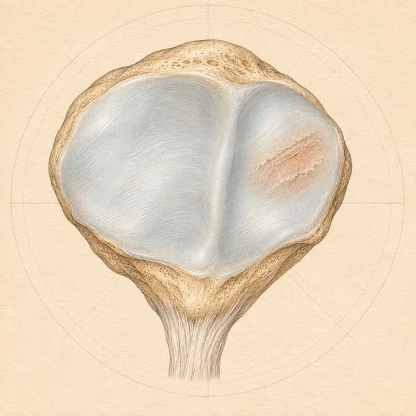
Wording about the two cartilage pads between the thigh bone and the shin bone.
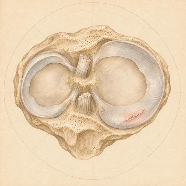
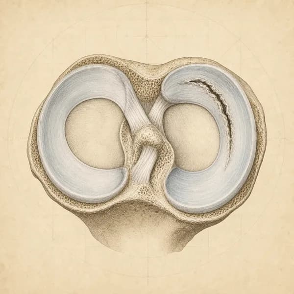
Grade 2 signal, degenerative, horizontal, complex and root tears, and extrusion on a knee MRI. How common tears are in painless knees, what the trials found about surgery, and when a surgical opinion is worth having.
Wording this guide explains- Degenerative tear
- Horizontal tear, horizontal cleavage
- Oblique or flap tear
- Vertical or longitudinal tear
- Radial tear
- Complex tear
- Bucket-handle tear
- Root tear
- Meniscal extrusion
- Parameniscal or meniscal cyst
Tendon and fat pad
Wording about the tendon below the kneecap and the fat pad behind it.
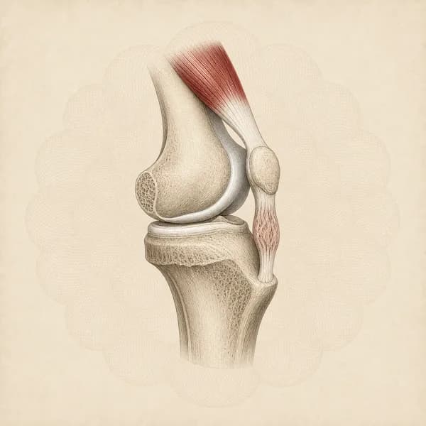
A thickened or changed tendon below the kneecap on an ultrasound or MRI, with the Doppler, partial tear and fat pad wording around it. Why it is common in painless jumping athletes and why a normal follow-up scan is not the goal.
Wording this guide explains- Patellar tendinosis or tendinopathy
- Mild proximal patellar tendinosis
- Thickening or increased thickness
- Hypoechoic area (ultrasound), increased signal (MRI)
- Increased Doppler signal, hyperemia, neovascularity
- Partial, intrasubstance or interstitial tear; fibre disruption
- Calcification
- Hoffa’s fat pad edema
- Infrapatellar bursitis, deep or superficial
- Sinding-Larsen-Johansson or Osgood-Schlatter
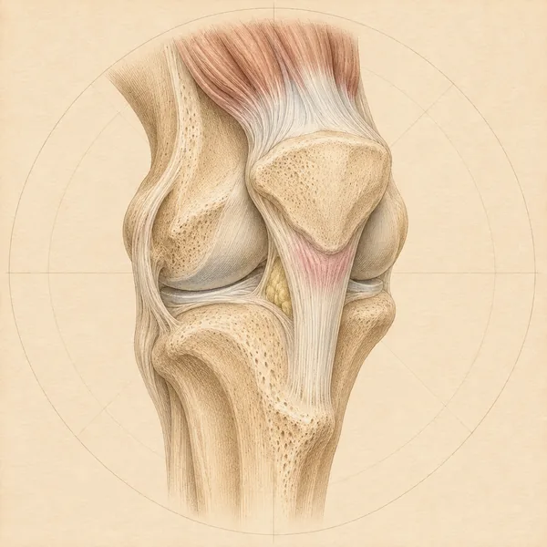
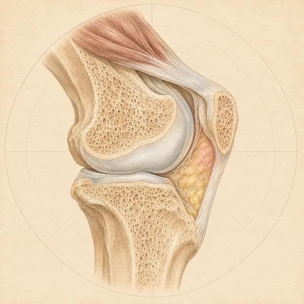
A bright patch in the fat pad behind the patellar tendon on a knee MRI, including superolateral edema, scarring after surgery and Hoffa-synovitis. Why it is common in painless knees and how fat pad pain differs from tendon and kneecap pain.
Wording this guide explains- Hoffa’s fat pad, infrapatellar fat pad, IFP
- Edema, high or increased signal, hyperintensity
- Superolateral Hoffa’s fat pad edema
- Patellar tendon to lateral femoral condyle friction, fat pad impingement
- Hoffa’s disease, Hoffa’s fat pad syndrome, fat pad syndrome
- Fibrosis, scarring, scar tissue, postoperative change
- Hoffa-synovitis
- Patella alta, high-riding patella, Insall-Salvati ratio
- TT-TG distance, lateral patellar displacement or tilt, trochlear dysplasia
- Mass, nodule, cyst or ganglion in the fat pad
Ligaments: ACL, MCL, LCL and PCL
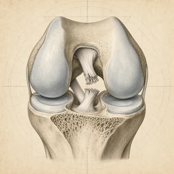
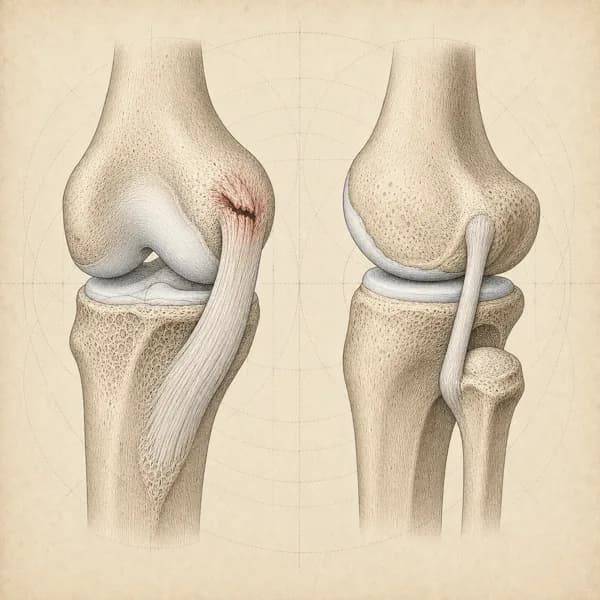
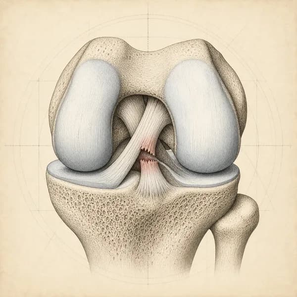
Ligament findings do not have a report guide yet. Each condition page covers what the injury means and how it is managed.
Anterior cruciate ligament tears, conservative and post-surgical rehab
Read the condition pageInner (MCL) and outer (LCL) knee ligament sprains and how they differ
Read the condition pagePosterior cruciate ligament tears, conservative and post-surgical rehab
Read the condition page
Related knee conditions
The condition pages behind the findings above: symptoms, treatment and recovery time.
Access, hours, and how to book
I see patients for knee pain at Endorphins Health & Wellness Centre in Burlington. The clinic serves people coming in from Burlington, Waterdown, Oakville, Hamilton, Flamborough, and Carlisle, with free parking on site and a ground-floor entrance.
- 4631 Palladium Way, Unit 6Burlington, ON L7M 0W9
- (905) 634-6000Direct insurance billing available. No physician referral needed.
Burlington hours
- Monday
- 1:30 to 7:00 PM
- Tuesday
- 3:30 to 8:00 PM
- Thursday
- 1:30 to 8:00 PM
- Wednesday
- 2:00 to 7:00 PM
- Friday
- 1:00 to 7:00 PM
- Tuesday
- 11:00 AM to 2:30 PM
- Saturday
- 12:00 to 3:00 PM