Deep Gluteal Syndrome
Sciatic nerve entrapment in deep gluteal space
Overview
The Science of Deep Gluteal Syndrome
Link copiedDeep Gluteal Syndrome (DGS) is a comprehensive term that describes the entrapment or irritation of the not just by the , but by a number of other structures in the deep buttock space, such as fibrous bands, the gemelli-obturator internus muscle group, or hamstring issues. The most common misconception is that all buttock and leg pain is "sciatica" coming from the . For decades, a condition called "Piriformis Syndrome" was used as a catch-all term for this type of pain. While the piriformis muscle can be involved, we now understand the situation is more complex.
The deep gluteal space is a busy anatomical neighborhood. The sciatic nerve must navigate a narrow tunnel surrounded by several deep hip rotator muscles like the piriformis. Irritation of the nerve in this space can cause DGS. This is the central mystery of DGS: buttock and leg pain that mimics a classic "pinched nerve" from the back, but originates from a completely different location - your back may be completely innocent.
The way you move can contribute to DGS. A gait pattern where the knee collapses inwards () can cause over-activity and eventual tightness of the deep external rotator muscles of the hip (like the piriformis) as they work overtime to try to control the femur. This tightness can contribute to nerve compression. Similarly, weakness in the or maximus can lead to compensatory strategies that overload these deeper muscles.
Living with nerve pain is unsettling. The tingling, burning, and unpredictable nature of the symptoms can create a high level of anxiety and fear. Patients often worry they have a serious spinal condition. Understanding that the nerve is simply "irritated" or "compressed" in the buttock, and not "damaged" in the spine, can significantly reduce fear.
Overview
Contributing Factors
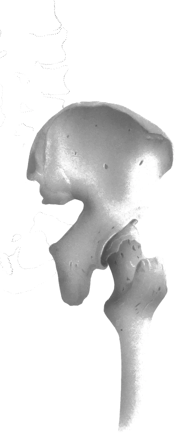
Link copiedThe deep gluteal space represents an anatomically constrained tunnel through which the must pass. This space is bounded by the greater sciatic notch superiorly, the inferiorly, the hip joint capsule anteriorly, and the gluteus maximus muscle posteriorly. Within this relatively small space, the sciatic nerve travels alongside or through several deep hip rotator muscles including the , superior and inferior gemelli, obturator internus, and quadratus femoris. Any factor that reduces the available space or increases muscle volume within this tunnel can compress the nerve.
Hip positioning dramatically influences the dimensions of the deep gluteal space. Cadaveric and imaging research demonstrates that hip flexion combined with and internal rotation - a position commonly assumed during sitting - reduces the available space for the sciatic nerve. This explains why prolonged sitting, particularly on hard surfaces or while driving, frequently aggravates symptoms. Each time you sit, particularly in a slouched posture with the hip flexed beyond 90 degrees, you mechanically narrow the tunnel through which your sciatic nerve travels.
The piriformis muscle, which runs from the sacrum to the , exhibits variable anatomy in its relationship to the sciatic nerve. In approximately 85% of individuals, the sciatic nerve exits the pelvis below the piriformis muscle. However, in about 15% of people, the nerve may pass through or above the piriformis, creating an anatomical predisposition to compression. When the piriformis contracts or increases in volume due to hypertrophy, spasm, or inflammation, it can compress the nerve against surrounding bony structures. Piriformis muscle contraction can compress the sciatic nerve against surrounding structures, and sustained compression above the threshold for neural ischemia can impair its blood supply.
Activity-related muscle hypertrophy plays a significant role in deep gluteal syndrome development. Athletes who perform repetitive hip external rotation activities - such as ballet dancers, soccer players, and ice skaters - develop significant piriformis and deep rotator muscle hypertrophy. Repeated demand on the deep external rotators may alter their bulk and tone over time, which can affect the space available for the sciatic nerve. This activity-induced hypertrophy explains why deep gluteal syndrome shows higher prevalence in certain athletic populations.
Sitting mechanics create sustained compression that differentiates deep gluteal syndrome from other causes of sciatic nerve pain. When you sit, your body weight compresses the soft tissues of the buttock between the ischial tuberosity (sitting bone) and the seat surface. This compression can reach pressures sufficient to impede venous return and create a mechanical load on the sciatic nerve. Sitting on hard surfaces generates higher peak pressures over the ischial tuberosity than sitting on cushioned surfaces, which is why cushioning and frequent position changes often ease symptoms.
Compensatory movement patterns contribute to deep gluteal syndrome through mechanisms involving abnormal muscle recruitment. When your or maximus muscles are weak or inhibited, the deeper external rotator muscles must work harder to stabilize the hip during activities like walking and running. This chronic overwork leads to muscle hypertrophy, increased muscle tone, and eventual nerve compression. When the larger gluteal muscles underperform, the deep external rotators tend to work harder to stabilise the hip through stance, increasing their tone and the compressive load they place on the nerve.
Prolonged nerve compression creates a cascade of pathophysiological changes beyond simple mechanical pressure. When compression exceeds 20-30 mmHg, it impedes intraneural blood flow, creating local ischemia. This triggers inflammation within the nerve itself, causing intraneural edema that further reduces the available space within the deep gluteal tunnel - creating a self-perpetuating cycle. On ultrasound, the symptomatic sciatic nerve can appear swollen in the deep gluteal space compared to the contralateral asymptomatic side, reflecting this inflammatory swelling.
Symptoms
Clinical Presentation
Link copiedPrimary Symptoms
Associated Symptoms
Typical pattern
The patient with Deep Gluteal Syndrome comes in with a story that is often a diagnostic puzzle. They describe a deep, aching, and sometimes burning or tingling pain in the buttock. Unlike simple muscle pain, this discomfort often travels, running down the back of their thigh, occasionally even into the lower leg. The symptoms can be vague and unpredictable. Sitting is a major aggravator, especially on hard surfaces or while driving, as it feels like they are putting pressure directly on a 'hot spot.' They might tell me, 'It feels like sciatica, but my back feels fine.' This is the central mystery of DGS: buttock and leg pain that mimics a classic 'pinched nerve' from the back, but originates from a completely different location.
Symptoms
Differential Diagnosis
Link copiedConditions with similar presentations:
Lumbar Radiculopathy
Key differences: Spinal signs, distribution, positive
Proximal Hamstring Tendinopathy
Key differences: Localized tenderness, sitting pain
Sacroiliac Joint Dysfunction
Key differences: tenderness, positive SI provocation tests
When to seek professional help
Research
Key Research & Evidence
Peer-reviewed studies supporting the treatment approach.
Finding
High diagnostic accuracy with combined clinical tests
Research details
Combination of seated piriformis stretch test with piriformis active test demonstrates sensitivity of 91% and specificity of 80% for endoscopic finding of sciatic nerve entrapment in deep gluteal space, providing reliable clinical diagnosis without imaging
Clinical relevance
Supports clinical diagnosis based on physical examination findings rather than requiring expensive imaging for initial assessment and treatment planning
Finding
Physiotherapy recommended as first-line treatment
Research details
Clinical guidelines recommend physiotherapy as first-line management following general guidelines on back pain and sciatica, with surgery considered only after failed conservative management in 50% of studies, though limited information details successful conservative management outcomes
Clinical relevance
Evidence supports conservative trial for 6-12 weeks before considering surgical options, though more research needed on specific physiotherapy protocols and outcomes
Finding
Condition affects 6-17% of secondary care sciatica patients
Research details
Between 6% and 17% of patients with sciatica presenting to secondary care meet diagnostic criteria for deep gluteal syndrome, indicating it represents significant proportion of non-discogenic sciatica cases often overlooked in clinical practice
Clinical relevance
Highlights importance of considering deep gluteal syndrome in differential diagnosis for patients with sciatica symptoms who don't demonstrate clear disc pathology on imaging
Management
Evidence-Based Management
Treatment strategies with the strongest support in the current literature.
Primary approach
combined with deep muscle release and strengthening achieves symptom resolution in 70-85% of deep gluteal syndrome cases by reducing nerve compression and improving function
Complementary
Activity modification and postural correction address contributing factors while gentle sustained stretching reduces tension in the and deep rotator muscles
Prevention & long-term
Sitting breaks and regular hip mobility reduce the likelihood of deep gluteal irritation in desk workers
Detailed management strategies
Avoid Prolonged Sitting
Reduces compression on the in the gluteal region
Important precautions
- Take frequent breaks
- Use cushioning
- Vary sitting positions
Gentle Neural Stretching
Maintains nerve mobility and reduces adhesions
Important precautions
- Avoid aggressive stretching
- Stop if symptoms worsen
Management
Treatment Techniques
Evidence-based manual therapy and intervention approaches.
Treatment approaches supported by current research and clinical guidelines
Recommended treatment approaches
Treatment approaches are individualized to each patient's needs and goals. All interventions require explicit informed consent, and treatment plans are collaboratively modified based on your preferences and response to care.
Dry Needling
Precise needle therapy targeting trigger points for effective pain relief and improved muscle function.
Soft Tissue & Myofascial Therapy
Targeted hands-on techniques to address muscle tension, pain, and movement restrictions.
Trigger Point Therapy
Focused pressure techniques to address painful trigger points and reduce muscle pain.
Rehabilitation
A Typical Rehabilitation Progression
Three phases, from settling symptoms to returning to full activity.
Recovery from Deep Gluteal Syndrome is usually staged: calm the symptoms first, then rebuild the strength and capacity of the area, then return to your full activities. The three phases below show the kind of progression the evidence supports and that I commonly work through in clinic. They are here to show you what the road can look like, not to act as a personal program.
- Phase 1
Desensitise the Nerve, Offload the Buttock (Weeks 1 to 4)
The early goal is calming irritability and the deep rotator muscle tone compressing it. Martin et al. (J Hip Preserv Surg 2015) describe the deep gluteal space as a tunnel whose dimensions are altered by hip position, which is why the earliest wins usually come from sitting and sleep set-up rather than from any one exercise.
Examples, not a prescription
- Sciatic in supine or long-sitting, 10 to 15 slow repetitions, stopping short of any leg symptom reproduction
- Gentle supine figure-4 positional hold at the first sense of stretch, 30 to 45 seconds, only if pain-free
- Clamshell and side-lying with a light band, 2 sets of 10 to 12 per side
- Diaphragmatic breathing in hook-lying plus standing and walking breaks every 20 to 30 minutes
- Sitting modifications: firm wedge cushion, offload the sore side, no back-pocket wallet, no cross-legged sitting
Ready to progress when
Leg symptoms stay at or above mid-thigh, sitting for 30 minutes is tolerable, and deep gluteal palpation tenderness has dropped by roughly half.
- Phase 2
Strengthen the Hip, Progress Nerve Mobility (Weeks 4 to 12)
Deep gluteal symptoms typically sit on top of and maximus weakness that drives the deeper rotators to compensate. Phase two shares the load across the larger hip muscles and moves nerve work from sliders to gentle tensioners.
Examples, not a prescription
- Glute bridge progressing to single-leg bridge, 3 sets of 8 to 12
- Banded lateral walks and monster walks, 2 to 3 sets of 10 to 12 steps each direction
- Split squat and step-up variations with a level pelvis, 3 sets of 6 to 10 per side
- Short-range sciatic nerve tensioners in supine or slump position, 8 to 10 controlled reps
- Hip hinge progressions: kettlebell deadlift, then single-leg Romanian deadlift
Ready to progress when
Single-leg bridge and step-up without reproducing buttock or leg symptoms, an hour of sitting with a flare under 3 out of 10, and a light walking or cycling programme back in the week.
- Phase 3
Rebuild Capacity and Recurrence-Proof (Months 3 to 6)
The final phase restores capacity for running, sport, or demanding work and gives the patient a minimum maintenance dose. Deep gluteal syndrome comes back when hip loading drops off, so the exit plan matters as much as the acute rehab.
Examples, not a prescription
- Loaded hinges (trap-bar or conventional deadlift), 3 to 4 sets of 3 to 6 at working load
- Front squat or rear-foot-elevated split squat at working loads
- Graded walk-run progression on flat terrain before hills or speed
- Low pogo hops progressing to lateral bounds for field-sport athletes
- Twice-weekly hip and trunk maintenance the patient will sustain independently
Ready to progress when
Full return to sport and work demands, independent flare management, and a written weekly plan the patient will actually do.
Management
Prognosis & Recovery
What outcomes and recovery factors typically look like.
Expected timeline
Conservative management typically trialed for 6-12 weeks, with many patients experiencing improvement
Natural history
Generally responds well to conservative management when properly diagnosed. Chronic cases may require more intensive intervention
Factors affecting recovery
Management
Measuring Progress
How to track the recovery arc week to week.
Day-to-day tracking
I track what changes day to day: pain interference with key tasks, movement quality during functional tests, and your confidence with daily activities
Assessment tools
Condition-specific questionnaires when useful (like the Oswestry for back pain or DASH for shoulder conditions)
Activity targets
One activity target that matches your goal - whether that's returning to sport, work tasks, or daily activities without limitation
Management
Frequently Asked Questions
Common concerns and answers about this condition.
Is this sciatica from my back?
Is this sciatica from my back?
Not usually, and that is the point of the diagnosis. Classical sciatica from a disc reproduces with , slump testing, and often comes with back pain or a clear pattern. Deep gluteal syndrome reproduces with direct palpation in the buttock and with positions that load the in the deep gluteal space. Martin et al. (J Hip Preserv Surg 2015) framed it clearly: this is non-discogenic, extrapelvic sciatic nerve entrapment. Same nerve, different location.
Why does sitting make it so much worse?
Why does sitting make it so much worse?
Sitting compresses the soft tissues of the buttock between the sitting bone and the seat, and hip flexion combined with and internal rotation narrows the deep gluteal space itself. You have to sit badly to feel it, and most people sit exactly that way all day. For flares, I recommend standing breaks every 20 to 30 minutes and a firm wedge cushion that offloads the sore side.
Should I stretch my piriformis aggressively?
Should I stretch my piriformis aggressively?
No. Forcing end-range stretches on an already nerve often makes symptoms worse the next day. Gentle positional holds that do not reproduce leg symptoms, combined with and glute strengthening, settle this more reliably than repeatedly yanking the knee to the opposite shoulder.
Do I need an MRI?
Do I need an MRI?
Not for most cases. The diagnosis is clinical and relies on a negative exam combined with specific provocation of the deep gluteal space. MRI is reserved for red flags, failure to progress with conservative care, or when the differential remains unclear. Ultrasound with an experienced operator sometimes adds value for targeted injection planning.
How long does it take to settle?
How long does it take to settle?
Most cases settle meaningfully over 6 to 12 weeks, though a chronic presentation with deconditioned glutes can take longer. Duration before starting rehab is the biggest predictor of speed. Systematic reviews of surgical management (Kay et al., Arthroscopy 2017) reserve surgery for clearly identified structural entrapment after a fair conservative trial, which most people never need.
Can I keep running?
Can I keep running?
Often yes, at a modified dose, provided running does not reliably flare leg symptoms. Short, flat, easy-paced runs are usually tolerated before long runs, hills, or speed. The flare pattern I see most often is long sitting before or after a run, not the run itself.
What actually compresses the nerve if it is not always the piriformis?
What actually compresses the nerve if it is not always the piriformis?
The deep gluteal space has several possible culprits: the in some anatomical variants, fibrous bands, the gemelli-obturator internus group, the hamstring origin, and vascular structures. Hernando et al. (Skeletal Radiology 2015) mapped this thoroughly. Part of the assessment is working out which structure is most likely in your case so the loading plan targets the right one.
Related Conditions
Conditions I commonly see alongside, or confused with, this one.
- Anatomically related
Piriformis Syndrome
Piriformis syndrome is part of deep gluteal syndrome spectrum
- Shares symptoms
Sciatica
Both cause sciatic-type pain; DGS involves peripheral nerve entrapment
- Anatomically related
Lateral Hip Pain & Gluteal Tendinopathy
Both involve deep gluteal muscles and hip region