Metatarsalgia
Ball of foot pain, forefoot overload syndrome
Overview
The Science of Metatarsalgia
Link copiedMetatarsalgia represents a symptom complex rather than a specific diagnosis, describing pain under the in the . This mechanical overload syndrome occurs when weight-bearing forces exceed the tissues' adaptive capacity, leading to inflammation and pain in the plantar structures of the forefoot.
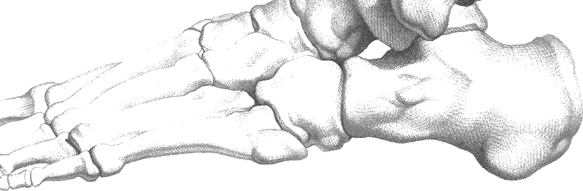
The normal forefoot functions as a complex lever system during push-off, with forces distributed across all five metatarsal heads. The central metatarsals (second and third) typically bear the greatest loads due to their length and position in the transverse arch. When this load distribution becomes pathological, excessive pressure concentrates under one or more metatarsal heads.
Several pathophysiological mechanisms contribute to metatarsalgia development. Primary metatarsalgia results from anatomical variants like elongated metatarsals or positioning that increase localized pressure. Secondary metatarsalgia develops from other conditions that alter forefoot mechanics, including , hammer toes, or first ray insufficiency that causes load transfer to the lesser metatarsals.
The plantar fat pad plays a crucial role in shock absorption and pressure distribution. With aging, this fat pad undergoes atrophy and displacement, reducing its protective effect. Repetitive loading can accelerate this process, particularly in high-impact activities or when combined with inappropriate footwear.
Inflammatory changes occur in response to excessive mechanical stress, affecting the joint capsules, surrounding soft tissues, and skin. Chronic inflammation can lead to synovitis of the joints, tears, and development of hyperkeratotic lesions (calluses) that further concentrate pressure points.
Overview
Contributing Factors
Link copiedNormal depend on coordinated function of the metatarsal parabola, transverse arch, and . During the propulsive phase of gait, the contact the ground sequentially, with forces distributed according to metatarsal length and position.
The first metatarsal typically bears 30-40% of forefoot load, while the lesser metatarsals share the remainder. This distribution depends on proper first ray function, adequate strength, and normal mechanics. When first ray insufficiency occurs (from hallux , , or functional weakness), load transfers excessively to the central metatarsals.
Transverse arch collapse represents a common biomechanical dysfunction contributing to metatarsalgia. As the arch flattens, the metatarsal heads spread apart and lose their coordinated load-sharing mechanism. This creates focal pressure points under individual metatarsal heads, particularly the second and third.
Calf muscle tightness significantly impacts forefoot loading by limiting ankle during midstance. This forces earlier heel rise and prolonged forefoot contact time, increasing the duration and magnitude of metatarsal loading. The resultant compensatory changes can overload the forefoot structures.
Footwear choices dramatically affect forefoot biomechanics. High heels shift body weight forward, increasing forefoot loading. Shoes with inadequate cushioning or support fail to attenuate impact forces, while narrow toe boxes compress the transverse arch and alter normal pressure distribution patterns.
muscle weakness contributes to metatarsalgia by reducing the foot's natural shock absorption capacity. These muscles help maintain the transverse arch and provide dynamic stabilization during loading. When weakened, they cannot effectively distribute forces, leading to concentrated pressure under individual metatarsal heads.
Symptoms
Clinical Presentation
Link copiedPrimary Symptoms
Associated Symptoms
Typical pattern
Pain typically begins gradually, initially occurring only during high-impact activities but progressing to affect normal walking. Symptoms worsen throughout the day with activity and improve with rest. Morning stiffness may be present, and pain often increases toward evening after daily activities.
Symptoms
Differential Diagnosis
Link copiedConditions with similar presentations:
Morton's Neuroma
Key differences: Neurological symptoms between toes, positive Mulder's sign, relief with toe manipulation
Stress Fracture of Metatarsal
Key differences: Point bone tenderness, positive percussion test, symptoms worsen with impact
Plantar Plate Tear
Key differences: Specific joint , positive drawer test, often involves second toe
Sesamoiditis
Key differences: Pain under first , worse with great toe extension
Metatarsophalangeal Joint Synovitis
Key differences: Joint-line tenderness, pain with passive motion, possible visible swelling
When to seek professional help
Research
Key Research & Evidence
Peer-reviewed studies supporting the treatment approach.
Espinosa N, Maceira E, Myerson MS · 2008
Current Concept Review: Metatarsalgia
Foot & Ankle International · n=Narrative concept review
Key findings
This concept review describes metatarsalgia as a symptom complex driven by abnormal forefoot load distribution and outlines conservative management, including footwear modification and orthotic offloading with metatarsal support, as the first-line approach before considering surgery.
Clinical relevance
Establishes evidence base for orthotic intervention as first-line treatment for metatarsalgia, supporting conservative management approach
Espinosa N, Maceira E, Myerson MS. Current Concept Review: Metatarsalgia. Foot Ankle Int. 2008;29(8):871-879.
Hastings MK, Mueller MJ, Pilgram TK, Lott DJ, Commean PK, Johnson JE · 2007
Effect of metatarsal pad placement on plantar pressure in people with diabetes mellitus and peripheral neuropathy
Foot & Ankle International · n=Plantar pressure measurement study
Key findings
Metatarsal pads positioned just proximal to the metatarsal head (about 6 to 11mm proximal) reduced peak forefoot pressure by an average of roughly one third, indicating that accurate pad placement proximal to the symptomatic head is what drives the offloading effect.
Clinical relevance
Provides biomechanical evidence that pad placement proximal to the metatarsal head reduces peak plantar pressure, supporting careful pad positioning in patient education
Hastings MK, Mueller MJ, Pilgram TK, Lott DJ, Commean PK, Johnson JE. Effect of metatarsal pad placement on plantar pressure in people with diabetes mellitus and peripheral neuropathy. Foot Ankle Int. 2007;28(1):84-88.
Chen WM, Lee SJ, Lee PVS · 2015
Plantar pressure relief under the metatarsal heads: therapeutic insole design using three-dimensional finite element model of the foot
Journal of Biomechanics · n=Finite element modelling study
Key findings
Computational modelling of therapeutic insoles found that a metatarsal pad combined with an arch support, rather than a flat insole, produced the greatest reduction in peak plantar pressure under the central metatarsal heads, reinforcing that offloading geometry matters more than cushioning alone.
Clinical relevance
Supports the use of contoured orthoses with metatarsal support for pressure redistribution, consistent with a conservative offloading approach to metatarsalgia
Chen WM, Lee SJ, Lee PVS. Plantar pressure relief under the metatarsal heads: therapeutic insole design using three-dimensional finite element model of the foot. J Biomech. 2015;48(4):659-665.
McKeon PO, Hertel J, Bramble D, Davis I · 2015
The foot core system: a new paradigm for understanding intrinsic foot muscle function
British Journal of Sports Medicine · n=Conceptual review
Key findings
This review proposes the foot core paradigm, describing how the intrinsic foot muscles support the arches and dynamically stabilise the foot during loading; the authors argue that intrinsic muscle function is often overlooked and can be targeted with specific strengthening to improve foot stability and load distribution.
Clinical relevance
Provides the conceptual basis for exercise interventions targeting intrinsic foot muscle function as an adjunct to orthotic offloading in forefoot pain
McKeon PO, Hertel J, Bramble D, Davis I. The foot core system: a new paradigm for understanding intrinsic foot muscle function. Br J Sports Med. 2015;49(5):290.
Management
Evidence-Based Management
Treatment strategies with the strongest support in the current literature.
Primary approach
Load redistribution through properly fitted orthotic devices with padding provides meaningful pain relief for many patients over several weeks of consistent use
Complementary
muscle strengthening combined with calf flexibility exercises addresses underlying biomechanical dysfunction that contributes to overload
Prevention & long-term
Appropriate footwear selection with adequate cushioning and gradual activity progression reduces the mechanical overload that drives most presentations in at-risk individuals
Detailed management strategies
Proper Load Redistribution Techniques
Metatarsal pads placed proximal to painful areas redistribute weight away from overloaded . Proper pad placement reduces peak pressure, leading to symptom improvement
Important precautions
- Pad placement is critical - too far forward increases pressure
- Start with softer materials before progressing to firmer supports
- Replace worn pads to maintain effectiveness
Intrinsic Foot Muscle Strengthening
Strengthening small foot muscles improves the foot's natural shock absorption and helps maintain transverse arch integrity. This reduces focal pressure points and improves overall mechanics
Important precautions
- Start with simple exercises like toe spreading and marble picking
- Progress intensity gradually based on tolerance
- Consistency more important than intensity
Appropriate Footwear Selection
Shoes with adequate cushioning, low heels, and sufficient width reduce mechanical stress on . Proper footwear can eliminate symptoms in mild cases
Important precautions
- Avoid high heels and narrow shoes during treatment
- Replace worn-out shoes promptly
- Consider professional fitting for optimal results
Activity Modification and Cross-Training
Temporary reduction of high-impact activities allows inflamed tissues to heal while alternative exercises maintain cardiovascular fitness and prevent deconditioning
Important precautions
- Modification should be temporary, not permanent avoidance
- Swimming and cycling are excellent alternatives
- Gradual return to high-impact activities as symptoms resolve
Calf Stretching and Ankle Mobility
Tight calf muscles increase loading by limiting ankle . Regular stretching reduces forefoot pressure and improves overall gait mechanics
Important precautions
- Stretch gently and consistently rather than aggressively
- Include both gastrocnemius and soleus stretches
- Maintain stretches for 30-60 seconds for optimal benefit
Management
Treatment Techniques
Evidence-based manual therapy and intervention approaches.
Treatment approaches supported by current research and clinical guidelines
Rehabilitation
A Typical Rehabilitation Progression
Three phases, from settling symptoms to returning to full activity.
Recovery from Metatarsalgia is usually staged: calm the symptoms first, then rebuild the strength and capacity of the area, then return to your full activities. The three phases below show the kind of progression the evidence supports and that I commonly work through in clinic. They are here to show you what the road can look like, not to act as a personal program.
- Phase 1
Offload and Settle (Weeks 1 to 4)
Take peak pressure off the sore and calm the surrounding tissue. Most of the early improvement comes from shoe changes, accurate metatarsal pad placement, and a reduction in time spent in aggravating footwear. Exercise in this phase is supportive and gentle.
Examples, not a prescription
- Metatarsal pad placed proximal to the tender metatarsal head, positioned initially in clinic then reviewed after a week for tolerance
- Daily wear of a shoe with adequate cushioning, a roomy toe box, and a heel under roughly 2 cm, during work and walking
- Gentle calf and soleus stretching, 30 seconds for 3 repetitions per side twice daily, to restore ankle and reduce forefoot loading time
- Toe splay and short-foot holds introduced seated, 10 slow repetitions with 5-second holds, to begin reawakening the
- Activity modification: reduce prolonged standing blocks, break up long walks, and add a cushioned insole or secondary pair of supportive shoes if switching occupations or surfaces during the day
Ready to progress when
Walking tolerance of 20 to 30 minutes in supportive footwear with the pad in place without escalating forefoot pain, reduction in end-of-day aching compared to baseline, and no need to remove shoes for relief during the work day for 7 consecutive days.
- Phase 2
Build Forefoot Capacity (Weeks 4 to 10)
Once the acute overload has settled, rebuild the intrinsic foot musculature and calf complex that share forefoot load. Stronger intrinsics maintain the transverse arch and help distribute pressure across all five metatarsal heads rather than funnelling it under the middle three. Pad and footwear strategy continues throughout.
Examples, not a prescription
- Short-foot exercise progressed from seated to standing, 3 sets of 10 with 10-second holds, performed without toe clawing
- Toe splay holds against a resistance band looped around the forefoot, 3 sets of 10 with 5-second holds
- Towel scrunches and single-toe lifts (raising the big toe while keeping the lesser toes down, then reversing), 2 sets of 15 per direction
- Single-leg calf raises with the heel tracking cleanly over the second toe, 3 sets of 12 to 15, progressed from floor to step
- Single-leg balance on firm ground progressing to a foam pad, 3 sets of 30 to 45 seconds, emphasising a stable, wide forefoot contact
Ready to progress when
A visible short-foot hold without toe clawing, single-leg calf raise for 15 clean reps without forefoot pain above 3 out of 10, and return to normal daily walking and standing volume in appropriate shoes without next-day flares.
- Phase 3
Return to Loading and Long-Term Strategy (Months 3+)
Rebuild tolerance for the activities that previously provoked symptoms, including longer walks, running where relevant, and standing-intensive work. The long-term strategy is informed footwear use paired with maintenance strengthening, rather than permanent avoidance.
Examples, not a prescription
- Walk-run progression for runners, using a 10 percent weekly cap on volume, with softer surfaces reintroduced first and speed work last
- Continued intrinsic foot and calf strengthening twice weekly as maintenance rather than daily
- progression from double-leg pogo hops to single-leg forefoot bounces on a soft surface, 3 sets of 10 to 20, for athletes
- Footwear audit every 6 to 12 months, including checking for midsole compression and whether the pad position still matches the current painful area
- Selective reintroduction of narrower or dressier shoes for shorter blocks, with recovery in wider, cushioned footwear rather than all-day wear
Ready to progress when
Return to desired walking, running, or occupational demands without ball-of-foot pain or next-day flares, comfortable tolerance of previously aggravating shoes for meaningful blocks, and a maintenance plan that the patient is willing to stick with.
Management
Prognosis & Recovery
What outcomes and recovery factors typically look like.
Expected timeline
Conservative treatment typically shows initial improvement within 4-6 weeks, with significant pain reduction achieved by 8-12 weeks. Complete resolution may take 3-6 months depending on underlying factors and compliance with management strategies
Natural history
Primary metatarsalgia often responds well to conservative management with proper load redistribution. Secondary metatarsalgia may require treatment of underlying conditions for optimal outcomes. Without intervention, symptoms typically worsen due to progressive tissue breakdown and development of compensatory deformities
Factors affecting recovery
Management
Measuring Progress
How to track the recovery arc week to week.
Day-to-day tracking
I monitor your pain levels during specific activities like walking and stair climbing, assess pressure point tenderness, measure walking tolerance before symptom onset, and track your ability to perform daily activities without discomfort
Assessment tools
Foot and Ankle Ability Measure (FAAM) for functional assessment, Manchester-Oxford Foot Questionnaire (MOXFQ) for quality of life impact, and Visual Analog Scales for activity-specific pain tracking
Activity targets
Return to desired activities including exercise, work tasks, and recreational pursuits without ball-of-foot pain or functional limitations
Management
Frequently Asked Questions
Common concerns and answers about this condition.
Is metatarsalgia just a fancy word for ball-of-foot pain?
Is metatarsalgia just a fancy word for ball-of-foot pain?
Yes. Metatarsalgia is an umbrella term for pain under the rather than a specific diagnosis. The clinically useful question is what is driving it. Espinosa and colleagues (JAAOS 2010) separate primary metatarsalgia, where the anatomy itself concentrates load under the lesser metatarsals, from secondary metatarsalgia, where something else (a bunion, a stiff great toe, a Morton's neuroma, a tear, a stress reaction) shifts load where it should not be. The label tells you where it hurts. The work is figuring out why.
Why does it feel worse at the end of the day?
Why does it feel worse at the end of the day?
loading is cumulative. Every step during a long standing or walking day sends multiples of body weight through the . With less fat pad cushioning or an anatomical variant concentrating pressure, the tissue runs out of buffer by late afternoon and the aching quality dominates. Taking the shoes off and the relief feels instant because the compression from the upper stops. That pattern, pain that tracks with time on feet and settles with unloading, is classic mechanical overload.
Where does the metatarsal pad actually go?
Where does the metatarsal pad actually go?
Behind the painful , not on top of it. The pad lifts the metatarsal shaft from underneath, which opens space between the heads and takes the peak pressure off the sore one. Placement is roughly 5 to 10 mm proximal to the tender spot. A pad that sits directly under the painful head tends to make things worse, which is the most common reason people try metatarsal pads, hate them, and give up. When positioning is right, the pad is usually tolerated well within a day or two.
Do I need custom orthotics or will over-the-counter work?
Do I need custom orthotics or will over-the-counter work?
For most people, a well-positioned pad added to a decent off-the-shelf insole is a reasonable starting point. Custom orthoses tend to be worth the investment when the foot shape is unusual, when there is a rigid deformity already shaping how load lands, or when basic pads and footwear changes have been given a fair trial and have not settled symptoms. I try the simpler version first and escalate only if it is not doing the job.
How do I tell this apart from Morton's neuroma?
How do I tell this apart from Morton's neuroma?
Metatarsalgia tends to feel bruised, deep, and aching under the ball of the foot, often with a callus building up over the painful head. Morton's neuroma tends to feel electric, burning, or like a pebble in the shoe, with tingling or numbness into adjacent toes. They can coexist. When both are present, treating the mechanical overload first often settles the nerve component too, because shrinking the load on the also reduces the squeeze on the nerve.
Can strengthening really change ball-of-foot pain?
Can strengthening really change ball-of-foot pain?
It can, but it works slowly and only pairs well with offloading. McKeon and colleagues (Br J Sports Med 2015) describe the as a core system for the , and weak intrinsics leave the transverse arch unsupported, concentrating load under the middle . Short-foot holds, toe splay work, and calf raise variations done consistently over two to three months change how the forefoot shares load. Strengthening on its own rarely fixes a genuinely mechanically overloaded forefoot, but combined with pad placement and footwear work, it is a reliable part of the picture.
When should I worry about a stress fracture rather than metatarsalgia?
When should I worry about a stress fracture rather than metatarsalgia?
A usually declares itself with point tenderness over the bone, not the soft tissue, and pain that does not ease promptly when weight is off the foot. Night ache, a sharper quality on percussion, and a clear training spike in the preceding weeks shift suspicion that way. Metatarsalgia from overload should ease meaningfully within a minute or two of sitting down. When it does not, when weight-bearing is painful from the first step regardless of time of day, imaging is worth getting rather than grinding through.
Related Conditions
Conditions I commonly see alongside, or confused with, this one.
- Shares symptoms
Morton's Neuroma
Both cause forefoot pain and can be confused diagnostically
- Biomechanically linked
Hallux Valgus (Bunions)
Bunions can cause transfer metatarsalgia due to altered weight distribution
- Biomechanically linked
Hallux Rigidus
Stiff great toe causes transfer of forces to other metatarsals