Bursitis / Tendinitis
Inflammatory shoulder conditions
Overview
The Science of Bursitis / Tendinitis
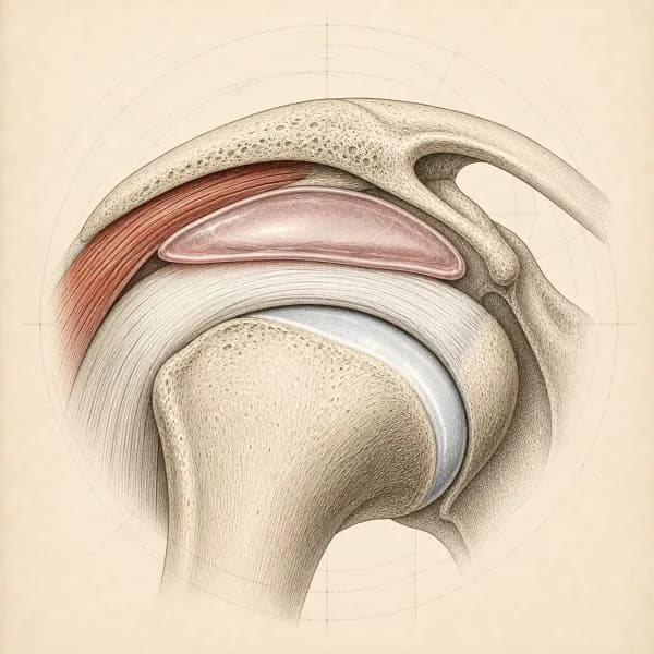
Link copiedShoulder , specifically bursitis, involves inflammation of the small fluid-filled sac () that sits between your tendons and the bony roof of your shoulder (). This bursa normally allows smooth gliding of the rotator cuff tendons beneath the acromion during arm movement.
When the subacromial space becomes narrowed due to , poor posture, muscle imbalances, or repetitive overhead activities, the bursa becomes compressed and irritated. This compression leads to inflammation, thickening of the bursa walls, and production of excess synovial fluid, creating a cycle of swelling and further compression.
The condition often coexists with rotator cuff and shoulder syndrome, as they share similar mechanical causes. The inflamed bursa can contribute to pain and dysfunction, but it's usually a secondary problem rather than the primary issue. Understanding this relationship is crucial because treating only the bursitis without addressing underlying mechanical problems often leads to recurrence.
Acute bursitis may result from direct trauma or sudden overuse, while chronic bursitis typically develops gradually from repetitive microtrauma and sustained mechanical irritation.
Overview
Contributing Factors
Link copiedThe space is a narrow area between your humeral head and the undersurface of the . During arm elevation, this space normally maintains about 6-14mm of clearance, but various factors can reduce this critical space.
Poor scapular mechanics significantly contribute to subacromial crowding. When your scapula doesn't rotate properly during arm elevation, it fails to maintain optimal clearance between the acromion and the underlying structures. This is often seen with weakness in the serratus anterior and lower trapezius muscles.
Forward head posture and rounded shoulders, common with extended desk or device use, alter the orientation of the acromion and reduce subacromial space. This postural pattern also changes the resting length and activation patterns of the muscles, making them less effective at maintaining proper humeral head position.
Rotator cuff weakness, particularly in the posterior and inferior aspects, allows superior migration of the humeral head during arm elevation. This upward translation narrows the subacromial space and increases compression forces on the and surrounding structures.
Symptoms
Clinical Presentation
Link copiedPrimary Symptoms
Associated Symptoms
Typical pattern
I typically see patients who describe a gradual onset of shoulder pain that started after a period of increased overhead activity or following a change in their usual routine. The pain is often worse at night and first thing in the morning, improving somewhat as they move around during the day.
Symptoms
Differential Diagnosis
Link copiedConditions with similar presentations:
Rotator Cuff Tendinopathy or Partial Tear
Key differences: Overlaps heavily with in the clinic, but weakness on resisted testing of a specific tendon points more toward tendon involvement. These frequently coexist and Lewis's related shoulder pain framework treats them as one continuum rather than separate labels.
Frozen Shoulder (Adhesive Capsulitis)
Key differences: Global loss of passive range of motion, especially external rotation at the side, with a capsular . typically preserves passive range and presents with a painful arc rather than global stiffness.
AC Joint Pathology
Key differences: Pain pinpointed at the top of the shoulder over the AC joint, tender on direct palpation, and worse with cross-body . presentations refer to the lateral deltoid rather than over the AC joint.
Calcific Tendinopathy
Key differences: Often a more acute onset with disproportionately severe pain, sometimes with visible calcium deposit on X-ray or ultrasound. Timeline and severity pattern differ from typical , though clinical tests overlap.
Cervical Radiculopathy (C5-C6)
Key differences: Arm symptoms in a pattern, positive Spurling's test, and pain reproduced by neck positioning rather than by direct shoulder loading. Passive shoulder range is preserved.
Biceps Tendinopathy
Key differences: Anterior shoulder pain localised over the , positive Speed and Yergason tests, and pain with resisted elbow flexion. Lateral deltoid referral from the cuff or is absent.
When to seek professional help
Research
Key Research & Evidence
Peer-reviewed studies supporting the treatment approach.
Finding
Physiotherapy shows lowest recurrence rates despite slower initial improvement
Research details
2023 randomized controlled trial with 111 patients compared corticosteroid injection (36 patients), physiotherapy (40 patients), and combined treatment (35 patients) over 8 weeks, finding physiotherapy group achieved 7.5% recurrence rate compared to 36.1% with injection alone and 17.1% with combined treatment, with statistically significant differences in shoulder flexion (p less than 0.003), pain (p less than 0.024), and external rotation (p less than 0.044)
Clinical relevance
Eight-week physiotherapy program provides superior long-term outcomes with substantially lower recurrence compared to corticosteroid injection, supporting physiotherapy as preferred first-line treatment despite corticosteroid providing faster short-term functional improvements
Finding
Scapular stabilization exercises reduce pain and improve function
Research details
2024 meta-analysis of 8 randomized controlled trials with 387 participants demonstrated scapular stabilization exercises produced weighted mean difference of minus 0.94 for pain on Visual Analog Scale (95% CI minus 1.23 to minus 0.65, p less than 0.001) and minus 10.10 for Shoulder Pain and Disability Index (95% CI minus 18.87 to minus 1.33, p equals 0.02)
Clinical relevance
Scapular stabilization exercises provide moderate evidence for pain reduction and functional improvement in subacromial pain syndrome, supporting their inclusion as core component of physiotherapy programs targeting scapular motor control
Finding
Ergonomic interventions reduce workplace-related shoulder pain
Research details
Meta-analysis of 24 randomized controlled trials with 4,086 workers showed ergonomic interventions reduced overall musculoskeletal pain with odds ratio of 0.64 (95% CI 0.56 to 0.73, p less than 0.00001) and upper back pain with odds ratio of 0.61 (95% CI 0.47 to 0.79, p equals 0.0002), with pain intensity decreasing by mean difference of minus 0.28 on Visual Analog Scale
Clinical relevance
Workplace ergonomic modifications combined with exercise programs significantly reduce shoulder and upper back pain in occupational settings, supporting multimodal approach for work-related subacromial conditions
Management
Evidence-Based Management
Treatment strategies with the strongest support in the current literature.
Primary approach
Conservative physiotherapy addressing underlying mechanical factors achieves significant pain reduction in 70-80% of shoulder cases by correcting scapular mechanics and function
Complementary
Anti-inflammatory modalities and activity modification provide symptom relief while corrective exercises address postural and biomechanical contributors
Prevention & long-term
Proper shoulder mechanics and regular strengthening reduce recurrence by maintaining space
Detailed management strategies
Posture Awareness and Correction
Maintaining proper shoulder blade position reduces crowding and prevents ongoing irritation of the
Important precautions
- Make gradual postural changes
- Set regular posture check reminders
Activity Modification
Temporarily avoiding or modifying overhead activities allows the inflamed to settle while maintaining overall shoulder function
Important precautions
- Don't completely stop moving your shoulder
- Gradually return to full activities
Gentle Range of Motion
Maintaining shoulder mobility prevents stiffness while avoiding positions that compress the inflamed
Important precautions
- Stay within comfortable range initially
- Avoid forceful stretching
Ice Application
Cold therapy can help reduce acute inflammation and provide temporary pain relief during flare-ups
Important precautions
- Limit to 15-20 minutes
- Use barrier to protect skin
Sleep Position Modification
Avoiding sleeping on the affected shoulder reduces direct pressure on the inflamed and improves sleep quality
Important precautions
- Use pillow support for positioning
- May take time to adjust to new positions
Management
Treatment Techniques
Evidence-based manual therapy and intervention approaches.
Treatment approaches supported by current research and clinical guidelines
Rehabilitation
A Typical Rehabilitation Progression
Three phases, from settling symptoms to returning to full activity.
Recovery from Bursitis is usually staged: calm the symptoms first, then rebuild the strength and capacity of the area, then return to your full activities. The three phases below show the kind of progression the evidence supports and that I commonly work through in clinic. They are here to show you what the road can look like, not to act as a personal program.
- Phase 1
Settle Irritability (Weeks 0 to 3)
Drop pain below the threshold where the and surrounding cuff can tolerate active loading. has evidence for short-term pain relief in and lets you produce force without the movement that typically aggravates pain. The 2022 JOSPT CPG supports active rehabilitation over rest from day one.
Examples, not a prescription
- Isometric external and internal rotation at the side, 4 to 5 holds of 30 to 45 seconds, daily
- Pain-free pendulum and small-arc active range of motion
- Scapular setting drills and supported low rows for posterior chain activation
- Sleep position modification and activity pacing education to reduce nightly flare-ups
- Relative rest from the specific provocative tasks (usually overhead reaching, lifting above shoulder height, sleeping on the affected side)
Ready to progress when
Night pain manageable, resting pain below 3/10, and isometric holds tolerated at 70% effort without a flare-up lasting beyond 24 hours.
- Phase 2
Restore Capacity (Weeks 3 to 10)
Build and scapular strength through progressively heavier resistance, which is the evidence-based driver of long-term improvement. Lewis's rotator cuff related shoulder pain framework and the 2022 JOSPT guideline both emphasise moving beyond light bands once irritability allows.
Examples, not a prescription
- Dumbbell or cable external rotation at the side, 3 sets of 8 to 12 reps with a controlled
- Side-lying external rotation progressing weekly by small dumbbell increments
- Prone Y, T, and W holds, and supported horizontal rows for scapular retraction
- Cable or band face pulls emphasising scapular retraction and posterior cuff activation
- Loaded carries (farmer and suitcase) to train shoulder girdle stability under load
Ready to progress when
Strength symmetry approaching 80% of the unaffected side on rotation testing, pain-free loading through full active range, and tolerance of moderate daily overhead reach.
- Phase 3
Return to Overhead and Sport (Months 2.5 to 5)
Restore capacity for overhead, ballistic, and end-range demands. Skipping this phase is why people plateau at partial recovery. Criterion-based rather than time-based progression matches the JOSPT guidance on rotator cuff rehabilitation.
Examples, not a prescription
- Overhead press progressions, starting with landmine press and advancing to full overhead dumbbell and barbell press
- Pulling work: lat pulldowns, progressions toward full pull-ups or assisted variations
- rotator cuff work (medicine ball chest pass, overhead throw, rebounder drills) for throwing athletes
- Sport or occupation-specific skill rehearsal with volume progression
- End-range strength work such as full overhead holds, Turkish get-ups, and bottoms-up kettlebell work for stability at length
Ready to progress when
Strength symmetry within 10% of the unaffected side across rotation, , and press testing, pain-free sport or occupation demands at expected volume, and confident overhead reaching in unplanned movements.
Management
Prognosis & Recovery
What outcomes and recovery factors typically look like.
Expected timeline
Most patients see improvement within 4-6 weeks with appropriate treatment. Full recovery typically takes 8-12 weeks depending on chronicity
Natural history
With appropriate mechanical correction, most cases resolve completely. Without addressing underlying causes, symptoms tend to recur
Factors affecting recovery
Management
Measuring Progress
How to track the recovery arc week to week.
Day-to-day tracking
I track your pain levels during specific activities like reaching overhead or sleeping, improvements in shoulder range of motion, and your ability to perform daily tasks without significant discomfort
Assessment tools
Shoulder Pain and Disability Index (SPADI) to monitor functional improvements and pain reduction over time
Activity targets
Return to full overhead activities and sleeping comfortably without recurring shoulder pain
Management
Frequently Asked Questions
Common concerns and answers about this condition.
Is shoulder bursitis actually its own condition?
Is shoulder bursitis actually its own condition?
Not really, most of the time. Isolated inflammation of the without involvement of the surrounding tendons is uncommon. Jeremy Lewis's 2016 paper on rotator cuff related shoulder pain and the 2022 JOSPT rotator cuff disorders clinical practice guideline both frame this as a continuum: the bursa, the tendon, and the capsule share one small space and get irritated together. When a report says , what it usually means is the whole subacromial complex is unhappy. This matters because treatment has to address the mechanics, not just the bursa.
Will a cortisone injection fix it?
Will a cortisone injection fix it?
It will probably help in the short term, and may not help in the long term. The Cochrane review of corticosteroid injections for shoulder pain found modest short-term benefit (roughly the first four to six weeks) compared to placebo, with limited evidence of benefit beyond that and no clear advantage over well-structured physiotherapy at three to six months. I do not object to injections when pain is disabling and someone cannot engage with exercise, but they are best used as a window to do the loading work, not a standalone solution. Injection is not something I do myself. When that route makes sense, I coordinate with a sports medicine physician who performs the procedure. A 2023 randomised trial in chronic (Hsieh et al., Clinical Rehabilitation) reported a substantially higher recurrence rate in the injection-only group (around 36 percent) than in the physiotherapy group (around 8 percent).
Why does my shoulder hurt more at night?
Why does my shoulder hurt more at night?
A few things stack up at night. Lying on the affected side compresses the and tendon, you lose the gravitational unloading that upright posture provides during the day, and blood flow to an already irritable area drops. I usually ask people to sleep on the opposite side with a pillow supporting the sore arm forward, or on their back with a small towel roll behind the scapula. Most people see a meaningful reduction in night pain within a couple of weeks.
Should I rest or keep moving?
Should I rest or keep moving?
Keep moving within reasonable limits. Complete rest tends to make shoulder pain worse because the quickly loses capacity and the joint stiffens. What I usually modify is the provocative activity (overhead work, reaching behind, sleeping position) rather than general movement. Light loading of the rotator cuff with isometrics and low-load external rotation is safe from the start for most presentations.
How long will this take to settle?
How long will this take to settle?
Realistic timelines are six to twelve weeks for meaningful improvement with a progressive programme. Around half of people are substantially better by six to eight weeks, and most of the rest continue to improve toward three months. Symptom duration before starting care is the biggest predictor of a slower recovery. Once pain has been present for over three months, I expect a longer road and set expectations accordingly.
Do I need imaging?
Do I need imaging?
Not for most presentations. Shoulder ultrasound and MRI pick up thickening, partial cuff tears, and shape changes in very high rates of pain-free shoulders, so they often add more uncertainty than clarity. I use imaging when I see suspicion of a significant tear, when there is red flag history like trauma with loss of active elevation, or when symptoms are not responding to six to eight weeks of appropriate rehab.
Is this going to turn into frozen shoulder?
Is this going to turn into frozen shoulder?
Not usually. Frozen shoulder has a distinct clinical picture (global loss of passive range of motion, particularly external rotation with the arm at the side) that is quite different from pain, which typically preserves passive range. That said, if someone guards their shoulder for months because of pain, secondary stiffness can creep in, which is another reason I prefer not to let people rest into complete immobility.
What exercises actually make a difference?
What exercises actually make a difference?
Three buckets, in this order: and low-load work at the side to reduce irritability, scapular control drills to restore the platform the shoulder needs, and then progressive loading into ranges and positions that matter for your life and sport. The 2022 JOSPT guideline and Lewis's rotator cuff related shoulder pain framework both support active, task-oriented rehabilitation over passive treatments. Details matter: sets, reps, tempo, and progression are what separate a programme that changes pain from one that just ticks boxes.
Related Conditions
Conditions I commonly see alongside, or confused with, this one.
- Common co-occurrence
Shoulder Impingement Syndrome
Subacromial impingement commonly causes secondary bursitis
- Shares symptoms
Rotator Cuff Injuries
Both cause shoulder pain and can coexist with similar presentations
- Shares symptoms
Frozen Shoulder
Both cause shoulder pain and stiffness with inflammatory components
- Anatomically related
Biceps Tendinopathy
Subacromial bursitis can affect nearby biceps tendon
- Shares symptoms
AC Joint Sprains
Both cause shoulder pain and can be confused diagnostically
Shoulder Instability / Dislocations
Getting back to it
Physiotherapy for bursitis / tendinitis, built around the activity you want back.