Sever's Disease
Calcaneal apophysitis, pediatric heel pain in growing athletes
Overview
The Science of Sever's Disease
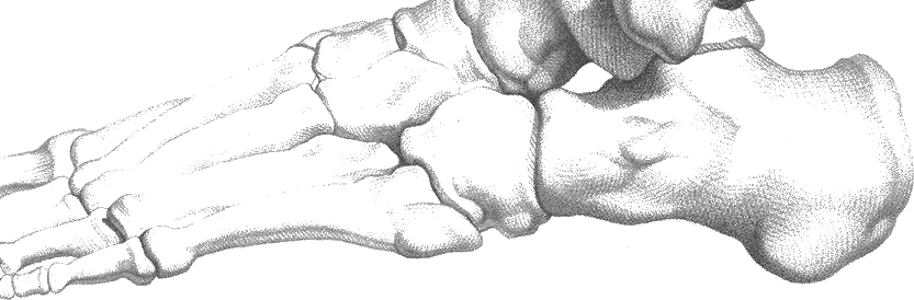
Link copiedSever's disease, formally known as , represents an inflammatory condition of the posterior calcaneal in skeletally immature children and adolescents. The calcaneal is a secondary that appears around age 8 and typically fuses with the main body of the calcaneus between ages 14-16 years.
During periods of rapid skeletal growth, the bones often grow faster than the surrounding soft tissues, creating increased tension in muscles and tendons. The Achilles tendon and both attach to the posterior aspect of the calcaneus, creating a traction force on the growth plate during activities involving running, jumping, or rapid direction changes.
The growth plate cartilage is inherently weaker than mature bone and more susceptible to stress-related injury. Repetitive traction forces from tight posterior muscle groups, combined with impact forces from athletic activities, create microtrauma within the growth plate. This leads to localized inflammation, increased blood flow, and pain characteristic of the condition.
The condition is essentially a stress reaction rather than an acute injury, developing gradually as cumulative stress exceeds the growth plate's adaptive capacity. Unlike adult , the problem lies within the bone itself rather than the tendon, explaining why rest is typically more effective than treatments targeting tendon .
The self-limiting nature of Sever's disease relates directly to skeletal maturation. As the growth plate closes and the apophysis fuses with the main calcaneal body, the weak link in the posterior heel complex is eliminated, and symptoms resolve permanently.
Overview
Contributing Factors
Link copiedThe posterior heel complex functions as an integrated system during weight-bearing activities, with forces transmitted from the calf muscles through the Achilles tendon to the insertion. In skeletally mature individuals, these forces are absorbed by mature bone tissue, but in growing children, the represents a point of relative weakness.
During the stance phase of gait, the gastrocnemius and soleus muscles contract to control forward progression of the tibia over the planted foot. This creates significant tension within the Achilles tendon, which translates to traction forces across the calcaneal . These forces are magnified during running and jumping activities where impact forces can exceed 3-5 times body weight.
Biomechanical factors that increase stress on the growth plate include excessive calf muscle tightness, which increases the baseline tension on the Achilles tendon throughout the gait cycle. Foot structure abnormalities such as pes planus (flat feet) or pes cavus (high arches) can alter the normal force distribution and increase stress concentration at the heel.
The also contributes to the biomechanical stress by creating a bowstring effect between the calcaneus and . During weight-bearing activities, tension in the plantar pulls on its calcaneal insertion, adding to the stress already created by Achilles tendon tension.
Poor footwear compounds the problem by failing to provide adequate shock absorption during heel strike, increasing the magnitude of impact forces transmitted through the growth plate. Hard playing surfaces similarly increase these impact forces, explaining why children playing on artificial turf or concrete surfaces often experience more severe symptoms.
Training errors, particularly rapid increases in activity intensity or duration, overwhelm the growth plate's adaptive capacity. The immature skeleton requires more gradual loading progressions compared to adult tissues, and violations of this principle commonly precipitate symptoms.
Symptoms
Clinical Presentation
Link copiedPrimary Symptoms
Associated Symptoms
Typical pattern
Pain typically follows a predictable pattern of activity-related onset, improvement with rest, and correlation with growth spurts. Symptoms are often worse at the beginning of sports seasons when activity levels increase rapidly. Morning symptoms are common but usually improve with gentle movement.
Symptoms
Differential Diagnosis
Link copiedConditions with similar presentations:
Calcaneal Stress Fracture
Key differences: More severe pain, often present at rest, positive imaging findings, typically in older adolescents
Plantar Fasciitis
Key differences: Pain primarily on plantar aspect of heel, worse with first steps in morning, uncommon in children under 10
Achilles Tendinopathy
Key differences: Pain and tenderness along the tendon itself rather than at bone insertion, more common in adolescents
Calcaneal Bursitis
Key differences: Swelling and tenderness over posterior heel prominence, associated with shoe friction
Tarsal Coalition
Key differences: Rigid flatfoot deformity, limited subtalar motion, pain with weight-bearing rather than just heel pain
When to seek professional help
Research
Key Research & Evidence
Peer-reviewed studies supporting the treatment approach.
Finding
Custom foot orthoses reduce pain by 68.6% and increase pressure pain threshold by 53.4%
Research details
A 2021 randomized controlled trial of 208 children aged 9-12 with calcaneal apophysitis found custom-made polypropylene foot orthoses produced a 68.6% reduction in VAS pain scores (95% CI: 74.5% to 62.7%) and 53.4% increase in pressure pain threshold (95% CI: 47.1% to 59.7%) compared to heel lifts over 12 weeks, with statistically significant improvements (p < 0.001)
Clinical relevance
Custom orthoses demonstrate superior outcomes compared to off-the-shelf heel lifts, supporting first-line treatment with properly fitted foot orthoses for pediatric patients with Sever's disease, with measurable improvements in both subjective pain and objective pressure tolerance
Finding
Conservative management can support return to sport within about two months
Research details
A 2025 review of 17 studies (Nweke, Cureus 2025) found that physical therapy facilitated return to sport within about two months when combined with activity modification and supportive measures
Clinical relevance
Conservative management with physical therapy and orthoses provides predictable recovery timelines for athletic populations, with treatment planning requiring consideration of bilateral involvement as a negative prognostic factor requiring extended rehabilitation duration
Research Database Expanding
Additional peer-reviewed studies are being reviewed and will be added to strengthen the evidence base for this condition.
Management
Evidence-Based Management
Treatment strategies with the strongest support in the current literature.
Primary approach
Activity modification combined with calf stretching programs is the mainstay of management, with symptoms typically resolving within 2-6 months and no recurrence after closure
Complementary
Heel padding and supportive footwear reduce impact forces and provide symptomatic relief while maintaining activity participation
Prevention & long-term
Gradual activity progression during growth periods and maintenance of calf flexibility may reduce the risk of symptoms in at-risk young athletes
Detailed management strategies
Smart Activity Modification and Cross-Training
Reducing high-impact activities while maintaining fitness through swimming, cycling, or other low-impact alternatives allows healing while preserving athletic conditioning
Important precautions
- Complete activity cessation rarely necessary and may lead to deconditioning
- Gradual return to impact activities as symptoms improve
- Monitor pain levels and adjust activities accordingly
Daily Calf Stretching Routine
Consistent stretching of both gastrocnemius and soleus muscles reduces tension on the and accelerates recovery while preventing recurrence
Important precautions
- Stretch should be comfortable, not painful
- Hold stretches for 30 seconds minimum
- Perform both straight-leg and bent-leg calf stretches daily
Proper Athletic Footwear with Heel Support
Shoes with adequate heel cushioning, arch support, and shock absorption reduce impact forces on the during activities
Important precautions
- Replace worn athletic shoes regularly
- Ensure proper fit with adequate heel and arch support
- Avoid worn-out or inappropriate footwear for sports
Heel Padding and Shock Absorption
Heel cups, gel pads, or cushioned insoles provide additional shock absorption and reduce direct pressure on the sensitive
Important precautions
- Ensure pads fit properly in shoes without creating tightness
- Replace worn padding regularly
- May need to size shoes slightly larger to accommodate padding
Ice Application After Aggravating Activities
Post-activity ice application reduces inflammation and pain, allowing continued participation in modified activities during the healing process
Important precautions
- Apply ice for 15-20 minutes maximum
- Use barrier between ice and skin
- Ice most effective when applied immediately after activity
Management
Treatment Techniques
Evidence-based manual therapy and intervention approaches.
Treatment approaches supported by current research and clinical guidelines
Rehabilitation
A Typical Rehabilitation Progression
Three phases, from settling symptoms to returning to full activity.
Recovery from Sever's Disease is usually staged: calm the symptoms first, then rebuild the strength and capacity of the area, then return to your full activities. The three phases below show the kind of progression the evidence supports and that I commonly work through in clinic. They are here to show you what the road can look like, not to act as a personal program.
- Phase 1
Settle the Heel (Weeks 1 to 3)
Calm the by reducing running and jumping volume, improving calf length, and cushioning heel strike. The aim is to keep the child in sport in some form while the settles.
Examples, not a prescription
- Gastrocnemius stretch against a wall, knee straight, 3 sets of 30 seconds, both sides, twice daily
- Soleus stretch against a wall, knee bent, 3 sets of 30 seconds, both sides, twice daily
- Heel cup or gel heel pad in both sport and school shoes
- Temporary reduction of running and jumping to roughly 50 percent of usual weekly volume, with cross-training like swimming or cycling to maintain fitness
- Ice for 10 to 15 minutes after any aggravating session
Ready to progress when
Heel squeeze tenderness clearly reduced, pain during normal walking at or under 2 out of 10, and no more than mild next-morning symptoms after modified training for 7 consecutive days.
- Phase 2
Rebuild Calf and Foot Capacity (Weeks 3 to 8)
Restore calf strength and foot control so the posterior chain can absorb running and jumping loads again. Calf flexibility work continues. Loading is introduced gradually and monitored using the 24-hour symptom response.
Examples, not a prescription
- Bilateral heel raise, 3 sets of 12 to 15, progressing to controlled tempo (2 seconds up, 2 seconds hold, 3 seconds down)
- Single-leg heel raise, building from 6 to 15 reps as tolerated
- Single-leg balance work (30 to 45 seconds per side), progressing to balance on foam or uneven surface
- Short-foot and toe-yoga exercises to build foot intrinsic capacity
- Gradual reintroduction of running, starting with run/walk intervals on soft surfaces
Ready to progress when
15 controlled single-leg heel raises without heel pain, calf flexibility within a few centimetres of the unaffected side on a weight-bearing lunge test, and pain-free jogging for 10 minutes on a supportive surface.
- Phase 3
Return to Sport with Load Management (Months 2 to 4)
Rebuild sport-specific demands (cutting, repeated jumping, sprinting) using graded volume increases. The goal is not just symptom freedom, it is teaching the child and family to recognize load spikes, growth spurts, and footwear issues that typically trigger recurrence.
Examples, not a prescription
- Bilateral jumping with soft landings, 2 sets of 10, progressing to single-leg hopping in straight lines
- Short acceleration drills (10 to 20 metres), progressing to change of direction work
- Progressive return to team training using a 10 to 15 percent weekly volume increase rule for running and jumping
- Full sport practice with one modified session per week during early return
- Ongoing calf flexibility and heel raise work 2 to 3 times per week as a maintenance habit
Ready to progress when
Two consecutive weeks of full team training and competition without heel pain, symmetrical calf strength (single-leg heel raise within 2 reps of the other side), and a family-level plan for managing the next growth spurt or season change.
Management
Prognosis & Recovery
What outcomes and recovery factors typically look like.
Expected timeline
Symptoms typically improve within 2-4 weeks of appropriate treatment and resolve completely within 2-6 months. Once fusion occurs (usually by age 16), symptoms do not recur
Natural history
Sever's disease is completely self-limiting and resolves with skeletal maturation. No long-term complications or recurrence occur once close. Most children return to full sports participation without restrictions
Factors affecting recovery
Management
Measuring Progress
How to track the recovery arc week to week.
Day-to-day tracking
I track your child's pain levels during different activities, monitor improvements in heel tenderness with squeeze testing, assess calf flexibility gains, and evaluate their ability to participate in sports and daily activities
Assessment tools
Pediatric Foot and Ankle Ability Measure (pFAAM), sport-specific return-to-play readiness scales, and parent-reported outcome measures for activity participation
Activity targets
Full return to desired sports and activities without heel pain, maintaining long-term habits of calf flexibility and appropriate training progression
Management
Frequently Asked Questions
Common concerns and answers about this condition.
Will my child grow out of Sever's disease?
Will my child grow out of Sever's disease?
Yes. Sever's is an of the , and once that growth plate fuses (typically around age 14 to 16), the vulnerable structure no longer exists. Most children return to full activity well before skeletal maturity, often within a few months of starting sensible management. It is a self-limiting condition, not a progressive injury.
Does my child need to stop sports completely?
Does my child need to stop sports completely?
Rarely. Complete rest deconditions the athlete and tends to delay return rather than help it. The usual plan is a temporary reduction in running and jumping volume, not removal. Most children can keep playing in some form while the heel settles, provided pain during and after activity stays manageable and settles overnight.
How long until the pain goes away?
How long until the pain goes away?
With activity modification, calf flexibility work, heel cups, and supportive footwear, most children feel clearly better within 2 to 4 weeks. Full resolution typically takes 2 to 6 months. Bilateral cases tend to take longer than one-sided cases. The condition does not recur once the has closed.
Why does the pain come back every season?
Why does the pain come back every season?
Sever's tends to flare when training load jumps quickly, like the start of a soccer season or a growth spurt combined with new activity. The common pattern I see is a child who felt fine all summer, starts three practices a week in September on turf, and has heel pain by week two. The fix is smoothing out those load spikes and maintaining calf flexibility year round rather than only when symptoms appear.
Do heel cups actually help?
Do heel cups actually help?
For many kids, yes. They cushion heel strike and can reduce pain enough to keep playing. A 2021 RCT in children with found custom foot orthoses outperformed simple heel lifts, but both reduced pain. I usually start with a good heel cup and supportive footwear before moving to custom orthotics, since the simple options are cheap and often enough.
Do I need an X-ray?
Do I need an X-ray?
Imaging is not usually needed to diagnose Sever's. It is a clinical diagnosis based on age, activity history, and a positive heel squeeze test. Imaging becomes relevant when the presentation is atypical, when pain is present at rest or at night, when there is a history of a specific injury, or when symptoms fail to improve with appropriate management over 8 to 12 weeks. In those cases I refer for imaging to rule out other causes.
What footwear helps most during a flare?
What footwear helps most during a flare?
Well-cushioned running shoes with a firm heel counter, replaced when they are visibly worn. Avoid flat, unsupportive shoes (including worn-out cleats and barefoot-style shoes) during a flare. On hard indoor court surfaces, adding a heel cup often makes a noticeable difference.
Related Conditions
Conditions I commonly see alongside, or confused with, this one.
- Anatomically related
Achilles Tendinopathy / Tendinitis
Both involve Achilles tendon attachment issues, though in different age groups
- Anatomically related
Growth Plate Injuries
Part of pediatric growth plate injury spectrum
- Anatomically related
Plantar Fasciitis & Heel Spurs
Both involve heel pain; plantar fascia attaches near calcaneal apophysis