Hallux Valgus (Bunions)
Big toe joint deformity, bunion pain and stiffness
Overview
The Science of Hallux Valgus (Bunions)
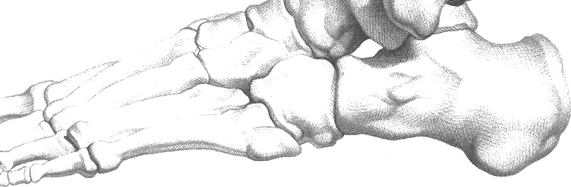
Link copiedrepresents a complex three-dimensional deformity of the first ray involving lateral deviation of the hallux at the , medial deviation of the first , and of the hallux. This progressive deformity results from a combination of intrinsic structural abnormalities and extrinsic environmental factors that disrupt the normal biomechanical balance of the first ray.
The deformity typically begins with attenuation of the medial joint capsule and stretching of the , allowing progressive lateral drift of the proximal phalanx. Simultaneously, the first metatarsal deviates medially (metatarsus primus ) due to the unopposed pull of the peroneus longus tendon and weakness of the tibialis anterior insertion.
As the deformity progresses, adaptive changes occur throughout the first ray. The complex becomes displaced laterally relative to the metatarsal head, creating a mechanical disadvantage for the flexor hallucis brevis and . The extensor hallucis longus and flexor hallucis longus tendons develop a bowstring effect, actually accelerating the deformity progression rather than providing corrective forces.
The overlying the medial eminence frequently becomes inflamed due to shoe pressure, creating the classic painful bunion presentation. Secondary arthritic changes develop within the metatarsophalangeal joint as the joint surfaces become incongruent. The altered mechanics also affect the entire , often leading to transfer metatarsalgia, lesser toe deformities, and compensatory gait changes that can affect the entire .
Overview
Contributing Factors
Link copiedNormal first ray function requires coordinated interaction between the first , proximal phalanx, complex, and surrounding musculature to provide stability during push-off and accommodate ground reaction forces during stance phase. The first must allow approximately 65-75 degrees of for normal gait mechanics.
In , several biomechanical factors contribute to deformity development and progression. Excessive foot increases the mobility of the first ray, allowing the first metatarsal to drift into under the influence of the peroneus longus muscle. This creates the characteristic intermetatarsal angle increase that defines the condition.
Restrictive footwear plays a crucial biomechanical role by forcing the hallux into a laterally deviated position repeatedly. High-heeled shoes compound this effect by increasing loading while simultaneously compressing the toes together in narrow toe boxes. This sustained positioning gradually overcomes the soft tissue constraints that normally maintain joint alignment.
The loss of the represents a critical biomechanical consequence of hallux valgus. As the great toe deviates laterally, its ability to tension the during push-off diminishes, reducing arch support and forcing the lesser metatarsals to accept greater loads. This load transfer often creates secondary pain under the second and third metatarsal heads.
Ground reaction force patterns change significantly with hallux valgus progression. Instead of the normal medial weight distribution pattern that utilizes the first ray for primary push-off power, patients develop lateral loading patterns that overload the lesser metatarsals and create inefficient propulsion mechanics.
Symptoms
Clinical Presentation
Link copiedPrimary Symptoms
Associated Symptoms
Typical pattern
Symptoms typically begin as mild shoe-related discomfort that progresses to constant pain. Deformity progression is generally slow but relentless without intervention. Pain patterns often correlate with footwear choices, activity level, and inflammatory episodes. Many patients experience symptom-free periods early in the condition's development.
Symptoms
Differential Diagnosis
Link copiedConditions with similar presentations:
Hallux Rigidus
Key differences: Loss of rather than lateral deviation; pain primarily with motion rather than shoe pressure
Sesamoiditis
Key differences: Pain primarily plantar to the great toe joint; tenderness over bones rather than medial eminence
First Metatarsophalangeal Joint Synovitis
Key differences: Acute onset joint swelling without progressive deformity; responds to anti-inflammatory treatment
Gout (First MTP Joint)
Key differences: Sudden onset severe pain with marked erythema; often associated with metabolic factors and acute attacks
Turf Toe (First MTP Sprain)
Key differences: Acute traumatic onset; pain with passive motion in all directions; history of hyperextension injury
When to seek professional help
Research
Key Research & Evidence
Peer-reviewed studies supporting the treatment approach.
Finding
Foot mobilization with exercise and toe separator reduces hallux valgus angle by 8.9° over 3 months with sustained 1-year results
Research details
Randomized clinical trial of 56 women receiving 36 sessions over 3 months showed hallux valgus angle decreased from 32.7° ± 4.2° to 23.8° ± 3.1° post-treatment (p<0.001), remaining stable at 25.8° ± 2.1° at 1-year follow-up (p<0.001). First-second intermetatarsal angle improved from 14° ± 1° to 11.8° ± 0.5° (p<0.001), maintained at 12° ± 0.9° at one year. Treatment included joint mobilization, strengthening exercises, and continuous toe separator use
Clinical relevance
Intensive multimodal conservative therapy combining mobilization, strengthening, and toe separator achieves clinically meaningful structural correction that persists long-term, offering evidence-based alternative to surgery for motivated patients with moderate hallux valgus
Finding
Toe-spread-out exercise combined with orthosis produces 3.41° angle reduction and 23.5% muscle hypertrophy in 8 weeks
Research details
Study of 24 subjects (19-29 years) performing 20 minutes daily, 4 days weekly for 8 weeks showed hallux valgus angle decreased from 18.33° to 14.92° (3.41° reduction) and abductor hallucis muscle cross-sectional area increased from 2.04 cm² to 2.52 cm² (0.48 cm² increase, 23.5% hypertrophy) in exercise group. During active abduction, angle decreased from 15.17° to 8.75° (6.42° reduction). Orthosis-only group showed no significant changes
Clinical relevance
Targeted intrinsic foot muscle strengthening drives both structural correction and muscle adaptation, with active exercise essential as orthosis alone produces no measurable improvement, emphasizing active rehabilitation over passive correction
Finding
Orthoses with toe separators are associated with a medium effect size for hallux valgus angle correction, though overall conservative efficacy is limited (Kwan et al., BMJ Open 2021)
Research details
2021 systematic review and meta-analysis of 9 studies found overall small effect size (SMD 0.31, 95% CI: 0.075 to 0.547) for hallux valgus angle reduction, with orthoses containing toe separator showing medium effect size (SMD 0.50, 95% CI: 0.189 to 0.803). Specific studies demonstrated angle reductions: Tang et al. 5.79°, Moulodi et al. 2.67°, Chadchavalpanichaya et al. 2.1°. 2-year prospective study found pain VAS scores decreased from median 52 at baseline to 21 at 12 months, maintained at 27 at 24 months (p<0.001)
Clinical relevance
While orthoses provide modest structural improvement and meaningful pain reduction sustained over 2 years, toe separator element is critical for optimal outcomes, and patients should maintain realistic expectations regarding degree of correction achievable with non-surgical management
Management
Evidence-Based Management
Treatment strategies with the strongest support in the current literature.
Primary approach
Conservative management combining footwear modification, padding, and orthotic therapy can relieve symptoms for many patients without halting deformity progression
Complementary
Targeted exercises and techniques help maintain joint mobility and muscle balance, potentially helping to slow progression when combined with other interventions
Prevention & long-term
Early intervention with footwear education and biomechanical assessment can meaningfully reduce the chance of symptomatic progression in at-risk individuals
Detailed management strategies
Proper Footwear Selection and Sizing
Shoes with wide toe boxes, low heels (<2cm), and soft uppers accommodate deformity and prevent pressure-related pain. Professional fitting ensures optimal comfort and function
Important precautions
- Measure feet regularly as size may change
- Shop for shoes in afternoon when feet are largest
- Prioritize width over length for proper fit
Bunion Protection and Padding Techniques
Protective pads over the bunion prominence and toe spacers between digits reduce friction forces and may provide temporary improvement in toe alignment
Important precautions
- Change pads regularly to prevent skin maceration
- Monitor for allergic reactions to adhesives
- Ensure pads don't create new pressure points
Daily Joint Mobility and Stretching Routine
Maintaining available great toe motion through gentle range of motion exercises and soft tissue stretching helps preserve function and may slow joint contracture development
Important precautions
- Avoid forceful manipulation that increases pain
- Perform exercises when joint is not acutely inflamed
- Focus on gentle, sustained stretches
Intrinsic Foot Muscle Strengthening
Strengthening the small muscles within the foot that help maintain arch structure and toe alignment may provide some resistance to deformity progression
Important precautions
- Progress exercises gradually to avoid muscle fatigue
- Maintain proper technique to avoid compensatory patterns
- Combine with other management strategies
Activity Modification and Load Management
Temporary reduction of high-impact activities and prolonged weight-bearing allows inflammation to settle while maintaining overall activity levels through appropriate modifications
Important precautions
- Modification should be temporary, not permanent activity cessation
- Find alternative activities that don't aggravate symptoms
- Gradually return to full activities as symptoms improve
Management
Treatment Techniques
Evidence-based manual therapy and intervention approaches.
Treatment approaches supported by current research and clinical guidelines
Rehabilitation
A Typical Rehabilitation Progression
Three phases, from settling symptoms to returning to full activity.
Recovery from Hallux Valgus is usually staged: calm the symptoms first, then rebuild the strength and capacity of the area, then return to your full activities. The three phases below show the kind of progression the evidence supports and that I commonly work through in clinic. They are here to show you what the road can look like, not to act as a personal program.
- Phase 1
Calm the Joint and Protect the Forefoot (Weeks 1 to 4)
Reduce the daily irritation at the bunion and the ball of the foot. Footwear swaps and a toe spacer tend to do most of the work here, with gentle mobility and intrinsic activation introduced alongside. Structural correction is not the goal of this phase. Symptom control and joint preservation are.
Examples, not a prescription
- Daily wear of a wide toe box shoe with a low heel, ideally with silicone toe spacer in situ for most of the day
- Big toe joint range of motion: gentle passive flexion and extension of the , 2 sets of 10 to 15 slow repetitions, twice daily, kept well within comfort
- hallucis activation seated, actively sliding the big toe away from the second toe without curling, 3 sets of 10 repetitions with 5-second holds
- Metatarsal pad placement behind the lesser if transfer pain under the ball of the foot is a significant feature
- Activity modification: reduce time in narrow or elevated shoes, break up long standing blocks, avoid bare feet on hard surfaces during flares
Ready to progress when
Bunion and pain reliably at 3 out of 10 or less during a normal day in the updated footwear, a visible and repeatable active big toe abduction contraction even if small, and no shoe-related flare that requires rest for 7 consecutive days.
- Phase 2
Intrinsic Foot Strengthening and First Ray Mobility (Weeks 4 to 12)
Rebuild the musculature, with a particular focus on abductor hallucis, and maintain first ray and big toe joint mobility. This is the phase where evidence for structural benefit is strongest, based on studies of toe-spread-out exercises and intensive multimodal conservative programs. Consistency matters more than intensity.
Examples, not a prescription
- Toe-spread-out exercise: seated then standing, actively spreading the toes apart including the big toe, 3 sets of 10 with 5-second holds, progressed to dynamic variations
- Short-foot holds, shortening the medial arch by drawing the ball of the foot back without curling the toes, 3 sets of 10 with 10-second holds
- Resisted abductor hallucis work using a band looped between the two big toes, pulling the hallux away from the midline, 3 sets of 15
- Big toe flexion strengthening with toes under a light resistance such as a folded towel, 3 sets of 15
- Self-mobilisation of the first into extension, sustained 20 to 30 second holds within comfort, performed before weight-bearing activity
- Single-leg calf raises with heel tracking over the second toe, 3 sets of 12 to 15, to reinforce first ray loading during push-off
Ready to progress when
A clearly visible active big toe abduction, short-foot hold for 10 seconds without toe clawing, big toe preserved near baseline, and normal daily walking volume in appropriate footwear without bunion or forefoot pain.
- Phase 3
Maintenance and Long-Term Progression Management (Months 3+)
Preserve the gains. Continue intrinsic strengthening at a maintenance dose and watch for early signs of progression, transfer pain, lesser toe drift, so they can be addressed before they become established. For people returning to running or higher-demand activities, rebuild tolerance with attention to forefoot mechanics.
Examples, not a prescription
- Intrinsic foot work 2 to 3 times weekly at maintenance dose rather than daily
- Continued toe spacer use during daily activity where tolerated
- Running or walking volume progressed using a 10 percent weekly rule, with attention to whether the big toe push-off is staying active
- Periodic footwear audit every 6 to 12 months to check width, heel height, and upper wear at the bunion
- Re-introduction of narrower or dressier footwear for shorter blocks, paired with recovery time in wide toe box shoes rather than all-day wear
- Early review with a foot and ankle surgeon if pain escalates meaningfully, transfer metatarsalgia becomes persistent, or lesser toe deformity is developing
Ready to progress when
Stable, comfortable function in desired daily activities, preserved big toe active abduction and joint mobility, and a maintenance routine the patient is realistically willing to sustain over the long term.
Management
Prognosis & Recovery
What outcomes and recovery factors typically look like.
Expected timeline
Conservative treatment provides meaningful symptom relief for many patients over the first few months, though deformity progression continues at variable rates. Long-term management required to maintain symptom control and function
Natural history
is typically progressive without intervention, with deformity angles tending to increase gradually over time. Symptom severity does not always correlate with deformity magnitude. Surgical intervention may become necessary in some cases when conservative management fails to maintain acceptable function and comfort
Factors affecting recovery
Management
Measuring Progress
How to track the recovery arc week to week.
Day-to-day tracking
I track your pain levels with different footwear and activities, measure available joint motion using standardized techniques, assess your comfort with recommended shoe modifications, and monitor any changes in deformity progression
Assessment tools
Manchester-Oxford Foot Questionnaire (MOXFQ) for symptom tracking, American Orthopaedic Foot and Ankle Society (AOFAS) Hallux Score for functional assessment, and patient-specific functional scale for activity goals
Activity targets
Maintain pain-free function for desired daily and recreational activities while slowing deformity progression and preventing development of secondary foot problems
Management
Frequently Asked Questions
Common concerns and answers about this condition.
Can physiotherapy actually straighten my bunion?
Can physiotherapy actually straighten my bunion?
No, and I would be honest with anyone who says otherwise. is a structural, three-dimensional deformity involving the first , the proximal phalanx, and the complex. Conservative care does not reverse that structure. What it can reliably do is reduce pain, improve how the shares load, maintain joint mobility, and slow progression. A Cochrane review of conservative interventions (Ferrari and colleagues) concluded that surgery tends to do better for pain than orthoses and exercise, but also that non-surgical measures remain a reasonable first line for people who are not ready for or not suitable for surgery.
So what is the point of exercise if it will not fix the bunion?
So what is the point of exercise if it will not fix the bunion?
The point is function, not cosmesis. Abdalbary and a handful of small trials have shown that strengthening, toe separator use, and first ray mobilisation can modestly reduce the angle and more reliably reduce pain over months. The bigger prize, though, is keeping the joint mobile, keeping the hallucis engaged, and preventing the secondary problems, transfer metatarsalgia, lesser toe deformities, compensatory gait changes, that tend to cause more disability than the bunion itself.
Are toe spacers and bunion splints worth using?
Are toe spacers and bunion splints worth using?
Toe spacers, used consistently, can reduce pain and improve the angle slightly while they are in place. Night splints have mixed evidence. They are not a substitute for active work, but they are a reasonable adjunct when the is being actively irritated or when worn alongside daily exercises. I prefer silicone spacers that can be worn with wide toe box shoes during the day, rather than rigid splints that only get used at night and skip the active loading piece.
What does the right shoe look like?
What does the right shoe look like?
Wide toe box so the is not being forced further into with every step, low heel so load is not multiplied, and a soft upper that does not grind on the bunion. The length of the shoe matters less than the width across the forefoot, which is why a lot of people are technically in the right size but in the wrong shape. A shoe that is quiet on the feet for a full day, with room for a toe spacer if desired, is doing most of the conservative work.
When is surgery actually the right call?
When is surgery actually the right call?
When pain is limiting daily life despite a genuine trial of conservative care, when the deformity is progressing fast enough that lesser toe deformities or transfer pain are appearing, or when the joint itself is becoming arthritic and motion is being lost. Bunion surgery has evolved considerably and outcomes for appropriately selected patients are generally good, but it is not a small procedure and recovery is measured in months. I stay neutral on the decision and focus on making sure the conservative column has been honestly tried before the surgical column is opened.
Will my bunion keep getting worse no matter what I do?
Will my bunion keep getting worse no matter what I do?
Most bunions are slowly progressive, but the rate varies widely between people and is not entirely fixed. Good footwear, consistent loading, and managing contributors like excessive can slow progression meaningfully. Genetics and first ray set the baseline, daily choices influence the trajectory. The expectation I set with people is that the clock cannot be stopped, but daily care can often buy years of comfortable function, which for many is enough.
Why does the ball of my foot hurt when the bunion is at the big toe?
Why does the ball of my foot hurt when the bunion is at the big toe?
Because the big toe is no longer doing its share of push-off. First ray insufficiency shifts load onto the lesser , and transfer metatarsalgia is one of the most common reasons a bunion that was merely cosmetic for years suddenly becomes functionally painful. Treating the bunion without addressing the load redistribution usually misses the actual driver of day-to-day pain.
Related Conditions
Conditions I commonly see alongside, or confused with, this one.
- Common co-occurrence
Metatarsalgia
Bunions commonly cause transfer metatarsalgia due to altered forefoot mechanics
- Biomechanically linked
Morton's Neuroma
Bunion deformity can contribute to forefoot nerve compression
- Common co-occurrence
Hammer Toe Deformities
Hallux valgus commonly leads to lesser toe deformities