Peroneal Tendinopathy
Lateral ankle tendon issues
Overview
The Science of Peroneal Tendinopathy
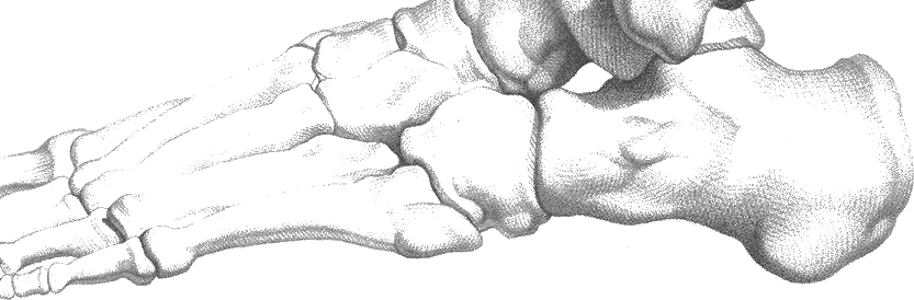
Link copiedPeroneal affects the fibularis longus and brevis tendons that run behind your lateral malleolus (outer ankle bone). These tendons are crucial for ankle stability, particularly during walking on uneven surfaces, and help prevent ankle sprains by providing lateral stability.
The peroneal tendons are subject to significant mechanical stress as they navigate around the sharp posterior edge of the fibula, held in place by the superior peroneal . This anatomical arrangement makes them vulnerable to friction and changes, particularly when the retinaculum is damaged or when there are underlying biomechanical issues.
Tendinopathy develops when the cumulative load on these tendons exceeds their adaptive capacity. This leads to a failed healing response characterized by disorganized , increased ground substance, and . The result is a painful, thickened tendon with reduced mechanical properties.
The condition often develops in conjunction with chronic ankle , where recurrent ankle sprains lead to peroneal muscle weakness and altered . Research shows that individuals with chronic ankle instability demonstrate reduced and delayed activation of peroneal muscles, creating a cycle where weak peroneals increase ankle instability, leading to further tendon stress and degeneration. This relationship helps explain why peroneal tendinopathy and chronic ankle instability so often occur together.
Overview
Contributing Factors
Link copiedYour peroneal muscles serve dual roles as foot everters and ankle lateral stabilizers. The fibularis longus also supports your medial longitudinal arch, while the fibularis brevis provides dynamic stability against ankle .
During the stance phase of walking, your peroneal muscles work eccentrically to control inversion and provide lateral stability. On uneven surfaces or during cutting movements in sports, these demands increase dramatically, making the tendons vulnerable to overuse injury.
The peroneal tendons must navigate a sharp turn around the lateral malleolus, similar to a rope moving around a pulley. This creates high friction forces, especially when ankle motion increases or when there are anatomical variations like a prominent peroneal tubercle.
When ankle is impaired following sprains, the peroneal muscles must work harder to provide conscious and subconscious stability. This increased demand, combined with potential weakness from previous injuries, creates the perfect environment for development.
Symptoms
Clinical Presentation
Link copiedPrimary Symptoms
Associated Symptoms
Typical pattern
I typically see patients with a history of ankle sprains who develop gradual onset lateral ankle pain. Many describe difficulty with activities like hiking on trails or sports requiring quick direction changes. The pain often improves with rest but returns quickly when activity resumes.
Symptoms
Differential Diagnosis
Link copiedConditions with similar presentations:
Chronic Lateral Ankle Ligament Injury or Instability
Key differences: Pain sits over the anterior talofibular or calcaneofibular ligaments at the outside of the joint, with a history of one clear sprain and a feeling that the ankle gives way on uneven ground. The peroneal tendons themselves feel comfortable on resisted , and tenderness does not track behind and below the lateral malleolus in the way does. The two often coexist, so the clinical question is which one is currently driving symptoms.
Peroneal Tendon Tear or Subluxation
Key differences: A palpable snap or a feeling of the tendons flicking over the back of the lateral malleolus during active foot points toward rather than straightforward . A sharp increase in pain with a specific cutting or landing event, persistent swelling, and a tendon that feels thickened with a focal divot on palpation raise concern for a split or partial tear and change the imaging threshold.
Lateral Ankle Bone Stress Injury
Key differences: Pain is reproduced with bony percussion or side-to-side compression of the fibula rather than with resisted . Weight-bearing pain persists regardless of warm-up, and night pain at rest is more common. Recent rapid training progression in a runner or hiker, or a stress riser like a single long day on rough terrain, raises suspicion.
Sinus Tarsi Syndrome
Key differences: Pain localises to the small depression just in front of the lateral malleolus, worst on uneven ground and when landing from a hop with the foot everted. The peroneal tendons themselves are non-tender and resisted is comfortable. A history of repeated ankle sprains with persistent lateral ankle discomfort is typical.
Sural Nerve Irritation
Key differences: The pain has a burning or tingling quality and follows the outer border of the foot rather than sitting in the tendon. Light percussion over the nerve behind the lateral malleolus reproduces symptoms, while resisted and direct tendon loading do not. Tight boot tops or a previous surgical scar are common contributors.
Cuboid Syndrome
Key differences: Deep pain on the outer midfoot rather than behind the lateral malleolus, often with a sense of something clunking or locking when pushing off. Cuboid-specific provocation such as direct plantar pressure under the cuboid reproduces the symptoms. Peroneal tendon palpation is usually comfortable, and resisted is not the primary reproducer.
When to seek professional help
Research
Key Research & Evidence
Peer-reviewed studies supporting the treatment approach.
Finding
Ultrasound-guided peroneal tendon sheath corticosteroid injection provided pain relief lasting more than one week in roughly 56% of patients
Research details
2019 study of 96 patients (109 injections) found 12.6% experienced 2-6 weeks relief, 6.9% had 7-12 weeks relief, and 36.8% achieved sustained relief beyond 12 weeks. Pre-injection symptom duration positively correlated with pain relief duration (p=0.036). Complication rate was low at 1.8%, with 25% of patients ultimately progressing to surgery
Clinical relevance
Ultrasound-guided injection represents a safe intermediate conservative option with low complication rates, particularly beneficial for patients with longer symptom duration who may achieve extended pain relief before considering surgical intervention
Research Database Expanding
Additional peer-reviewed studies are being reviewed and will be added to strengthen the evidence base for this condition.
Management
Evidence-Based Management
Treatment strategies with the strongest support in the current literature.
Primary approach
Combined progressive loading and ankle stability training can achieve good outcomes in peroneal , with addressing underlying chronic ankle being important for successful long-term outcomes
Complementary
strengthening promotes tendon remodeling while proprioceptive training addresses the neuromuscular deficits that contribute to both ankle and peroneal overload
Prevention & long-term
Early rehabilitation of ankle sprains and prevention of chronic ankle meaningfully reduces peroneal risk, as these conditions share common pathophysiological pathways
Detailed management strategies
Progressive Loading Program
Systematic loading helps tendons adapt and remodel while rebuilding strength and endurance of peroneal muscles
Important precautions
- Progress based on tendon response
- Some discomfort during exercise is acceptable
Balance and Proprioceptive Training
Improving ankle stability reduces excessive demands on peroneal tendons during functional activities
Important precautions
- Start with stable surfaces
- Progress to unstable surfaces gradually
Activity Modification
Temporarily reducing high-stress activities allows tendon healing while maintaining cardiovascular fitness
Important precautions
- Modify rather than completely avoid activity
- Use supportive footwear
Ankle Mobility Maintenance
Maintaining ankle range of motion prevents stiffness that could alter and increase tendon stress
Important precautions
- Gentle stretching within comfortable range
- Focus on all planes of movement
Management
Treatment Techniques
Evidence-based manual therapy and intervention approaches.
Treatment approaches supported by current research and clinical guidelines
Recommended treatment approaches
Treatment approaches are individualized to each patient's needs and goals. All interventions require explicit informed consent, and treatment plans are collaboratively modified based on your preferences and response to care.
Soft Tissue & Myofascial Therapy
Targeted hands-on techniques to address muscle tension, pain, and movement restrictions.
IASTM (Instrument Assisted Soft Tissue Mobilization)
Instrument-assisted techniques to address soft tissue restrictions and pain.
Rehabilitation
A Typical Rehabilitation Progression
Three phases, from settling symptoms to returning to full activity.
Recovery from Peroneal Tendinopathy is usually staged: calm the symptoms first, then rebuild the strength and capacity of the area, then return to your full activities. The three phases below show the kind of progression the evidence supports and that I commonly work through in clinic. They are here to show you what the road can look like, not to act as a personal program.
- Phase 1
Calming the Tendon and Reclaiming Balance (Weeks 1 to 4)
Bring day-to-day symptoms down to a workable level and begin to restore the peroneal activation that typically lags after sprain-related . is the anchor loading exercise, because it loads the tendon heavily without the repeated length changes that often provoke early irritation. Balance work rebuilds the proprioceptive input the lateral ankle has lost, given the delayed peroneal activation documented in chronic ankle instability research.
Examples, not a prescription
- Isometric eversion against a wall or a stable band, 5 sets of 45 seconds at a moderate effort, once daily
- Seated band eversion for isotonic tolerance, 3 sets of 12 to 15, slow tempo
- Single-leg balance on firm ground, eyes open, progressing to eyes closed, 3 sets of 30 seconds
- Calf raise variations on flat ground, 3 sets of 12, to share load with the triceps surae
- Activity modification: reduce uneven-terrain running and hill work by roughly 50 percent, keep volume on flatter surfaces
Ready to progress when
Pain during isometric eversion at or below 3 out of 10 for 7 consecutive days, single-leg balance on firm ground for 30 seconds without wobble, and no symptom rise 24 hours after loading sessions.
- Phase 2
Progressive Loading and Instability Challenge (Weeks 4 to 10)
Build peroneal strength through range and add graded instability. Progressive loading is the common thread across tendon rehab, and for this group it pairs naturally with the balance retraining that addresses the underlying chronic ankle instability picture. Some discomfort during loading is acceptable provided it settles within 24 hours and does not climb week over week.
Examples, not a prescription
- Heavy slow resistance eversion with a long band or cable, 3 sets of 6 to 10 reps, 3 seconds out and 3 seconds back, 3 times per week
- eversion with a manual overload, focusing on slow return from the everted position, 3 sets of 8 to 10
- Single-leg calf raise with the heel off a small step, 3 sets of 8 to 12, emphasising control on descent
- Single-leg balance on a foam pad or Airex, with reaches in multiple directions, 3 sets of 8 per direction per side
- Walking lunges and lateral step-ups to load the foot in functional positions, 3 sets of 8 to 10
Ready to progress when
Heavy slow resistance eversion tolerated at 3 out of 10 pain or less, single-leg heel raise at full range for 15 reps per side, stable balance on foam for 30 seconds with eyes closed, and two consecutive weeks of loading without a 24-hour flare.
- Phase 3
Return to Terrain, Cutting, and Sport (Months 3 to 6)
Restore the reactive stability the peroneal tendons are asked to deliver on uneven ground and in sport. Hopping, cutting, and landing rebuild the spring-like function the tendon needs, and trail or court surfaces are reintroduced gradually rather than in one jump. This phase also consolidates the instability work, since the highest recurrence risk sits in people who return to sport with the tendon settled but the ankle still underprepared.
Examples, not a prescription
- Double-leg pogo hops progressing to single-leg hops in place on firm ground, 3 sets of 15 to 20
- Lateral hop-and-stick onto the affected leg, 3 sets of 8, with a 2-second hold on landing
- Cutting drills starting at 45 degrees and progressing to 90, at submaximal then maximal intent
- Graded return to trail, hill, or uneven-surface training, using the 10 percent weekly volume rule
- Sport-specific reintegration, such as change of direction drills for soccer or court sports, in blocks with full recovery between
Ready to progress when
Single-leg hop symmetry within 10 percent of the unaffected side, return to previous training volume and terrain without next-day flares, FAAM Sport subscale trending toward pre-injury levels, and two consecutive weeks of full training without a symptom rise.
Management
Prognosis & Recovery
What outcomes and recovery factors typically look like.
Expected timeline
Symptoms often improve within 6-12 weeks with appropriate loading program, though complete resolution may take 3-4 months
Natural history
Most cases respond well to conservative management when underlying is addressed. Failure to improve may require surgical evaluation
Factors affecting recovery
Management
Measuring Progress
How to track the recovery arc week to week.
Day-to-day tracking
I monitor your pain during weight-bearing activities, improvements in single-leg balance, ankle stability during functional movements, and your ability to perform activities that previously caused symptoms
Assessment tools
Foot and Ankle Ability Measure (FAAM) and Victorian Institute of Sport Assessment-Achilles (VISA-A) adapted for peroneal tendons
Activity targets
Return to your desired activities including sports and uneven terrain walking without recurring lateral ankle pain
Management
Frequently Asked Questions
Common concerns and answers about this condition.
Why does the outside of my ankle hurt if I did not roll it recently?
Why does the outside of my ankle hurt if I did not roll it recently?
A history of rolled ankles years ago still matters. Once the lateral ligaments have been stretched, the peroneal tendons take on more of the stabilising work during walking and running, particularly on uneven ground. Over time that extra demand can overload the tendons even without a fresh sprain. This is why peroneal and chronic ankle so often travel together, and why rehab has to address both the tendon and the underlying instability rather than only the painful side.
How long does peroneal tendinopathy usually take to settle?
How long does peroneal tendinopathy usually take to settle?
Most people see meaningful change inside 8 to 12 weeks of consistent loading, with full return to running, hiking, or cutting sports often landing in the 3 to 6 month range. Cases that have been grumbling for over a year, or that have developed on a background of untreated ankle , take longer. The two biggest predictors of a faster timeline are catching it early and sticking with a progressive loading program rather than cycling through rest, flare, rest.
Should I rest completely, or keep moving?
Should I rest completely, or keep moving?
Complete rest is rarely the answer. The tendon needs load to remodel, and long periods off just delay the conversation. Most people do better on a modified plan: drop the aggravating volume by roughly a third, swap uneven-terrain running for flatter surfaces for a few weeks, and add a specific peroneal loading program twice weekly. The 24-hour rule is the gatekeeper. If pain during activity stays under about 5 out of 10 and settles within 24 hours, the dosage is acceptable.
Do orthotics help peroneal tendinopathy?
Do orthotics help peroneal tendinopathy?
They can, particularly when the foot sits in chronic or when an old cavus (high-arched) foot posture is overloading the lateral column. A lateral wedge or a lateral flare on the orthotic offloads the peroneals during stance. Orthotics are a support, not a cure. I use them as a bridge while strengthening rebuilds capacity, not as a long-term substitute for the loading program.
What is the difference between peroneal tendinopathy and an ankle sprain that just will not heal?
What is the difference between peroneal tendinopathy and an ankle sprain that just will not heal?
A typical lateral ankle sprain irritates the anterior talofibular ligament at the front of the joint, and symptoms usually centre on a specific spot just below the bony bump on the outside. Peroneal sits behind and below that bump, travels down the outer foot, and is reliably reproduced by resisted . Sprains that do not settle after 6 to 8 weeks often have a peroneal component that has been missed, and the rehab needs to shift toward tendon loading rather than early-sprain protocols.
Can I keep running with peroneal tendinopathy?
Can I keep running with peroneal tendinopathy?
Usually yes, with adjustments. Dropping weekly mileage by around a third, running on flatter surfaces temporarily, and spacing runs at least 48 hours apart gives the tendon time to adapt. Hills, trails, and speed work come back last. If the pain climbs above 5 out of 10 during runs, or morning stiffness starts lasting more than 15 minutes, the loading has outpaced the tendon and the plan needs to scale back for a week or two.
Is an ankle brace a good idea, or does it make me weaker?
Is an ankle brace a good idea, or does it make me weaker?
In the short term, a lace-up brace during higher-risk activity, such as trail runs, basketball, or hiking, reduces torque on the tendon and often allows training to continue. It does not weaken the foot provided rehab keeps building strength underneath it. The brace is a partner to the loading program, not a replacement. Most people phase it out once tolerance is back and balance work is consistent.
Related Conditions
Conditions I commonly see alongside, or confused with, this one.
- Common co-occurrence
Ankle Sprains
Chronic ankle instability leads to peroneal tendon overuse and tendinopathy
- Biomechanically linked
IT Band Syndrome
Both involve lateral structures; ankle dysfunction can affect entire lateral chain
- Biomechanically linked
Shin Splints
Both involve lower leg overuse; shared biomechanical factors in athletes