Hip Bursitis
Bursal inflammation causing localized hip pain
Overview
The Science of Hip Bursitis
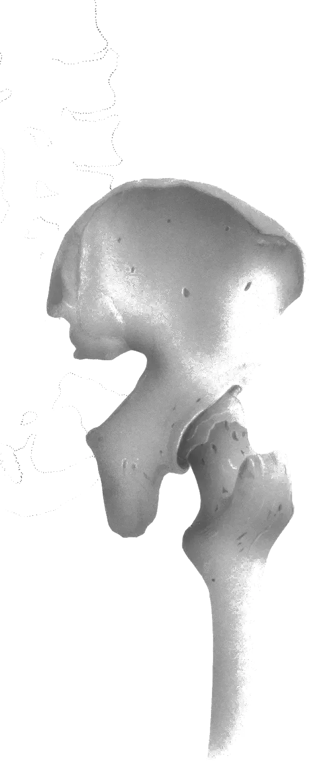
Link copiedHip involves inflammation of the fluid-filled sacs () that cushion the hip joint. The most commonly affected bursae are the bursa (lateral hip) and iliopsoas bursa (anterior hip). However, true isolated bursitis is actually less common than previously thought.
What was traditionally called "trochanteric bursitis" is now understood to be primarily Greater Trochanteric Pain Syndrome (GTPS) - a gluteal affecting the and minimus tendons. For years, lateral hip pain was attributed to an inflamed bursa, and the presumed treatment was rest, ice, and anti-inflammatory injections. Research has shown that the primary issue is frequently not an inflamed bursa, but a distressed gluteus medius or minimus tendon. The bursa can become secondarily irritated, but it's rarely the main driver.
This distinction is crucial because treating a tendinopathy is not about rest and inflammation control; it's about managing load and progressively strengthening the tendon. True isolated bursitis typically occurs secondary to other conditions, direct trauma, or in rare cases, infection or inflammatory conditions. The bursa becomes inflamed due to mechanical irritation or repetitive friction, but this is usually part of a broader mechanical problem rather than an isolated inflammatory condition.
Overview
Contributing Factors
Link copiedexist at locations where friction occurs between moving tissues - serving as fluid-filled cushions that reduce mechanical irritation. The bursa sits between the (IT) band and the greater trochanter bone, while the iliopsoas bursa sits between the iliopsoas tendon and the hip joint capsule or bony pelvis. Under normal circumstances, these bursae allow smooth gliding of these structures during hip movement. Inflammation develops when repetitive or excessive friction overwhelms the bursa's protective capacity.
For trochanteric , the mechanical problem involves the same compression forces discussed in Greater Trochanteric Pain Syndrome (GTPS). When your hip moves into adduction - bringing your thigh across your body's midline - the IT band tightens and compresses the trochanteric bursa against the greater trochanter. Positions that commonly create this compression include standing with weight shifted to one side, crossing your legs, and side-lying sleep postures where the top leg falls forward. substantially increases compressive load on the trochanteric bursa compared to neutral hip alignment.
Repetitive activities that involve and adduction cycles create a "bow-stringing" effect where the IT band repeatedly slides over the greater trochanter, generating friction forces on the underlying bursa. Running, particularly on banked surfaces where one hip experiences more adduction than the other, creates thousands of friction cycles per mile. Repeated stance-phase loading generates shear and friction forces over the greater trochanter that can accumulate over time to irritate the underlying bursa.
The iliopsoas bursa experiences different mechanical stresses. This bursa sits at the front of the hip where the iliopsoas tendon crosses the brim of the pelvis and hip joint capsule. During hip flexion and extension movements - such as running, climbing stairs, or performing sit-ups - the iliopsoas tendon slides back and forth across the bursa. In individuals with tight hip flexors or those performing high volumes of hip flexion activities, this repetitive motion generates friction that can inflame the bursa. Runners performing high weekly mileage with inadequate hip flexor flexibility are at greater risk of iliopsoas bursa irritation than recreational runners.
Body positioning during sleep creates sustained compression on hip bursae that prevents overnight recovery. When lying on your side, direct pressure compresses the trochanteric bursa for hours at a time. This sustained compression impedes blood flow to the bursal tissues and prevents the normal inflammatory healing processes from occurring during sleep. Clinically, sustained side-lying compression on the affected hip is a recognised aggravating factor, with the mechanical stress of prolonged compression impeding recovery.
Muscle weakness, particularly of the hip abductors, creates abnormal loading patterns that increase friction forces on the bursae. When your is weak, the IT band and tensor fasciae latae must work harder to stabilize the pelvis during single-leg stance. This increased muscle tension translates to greater compression and friction forces on the trochanteric bursa. Hip abductor weakness is associated with increased IT band tension during walking compared to normal strength, contributing to greater bursal compression.
Symptoms
Clinical Presentation
Link copiedPrimary Symptoms
Associated Symptoms
Typical pattern
Often develops following trauma, overuse, or as secondary to other hip conditions. presents with lateral hip pain, while iliopsoas bursitis causes anterior hip/groin pain.
Symptoms
Differential Diagnosis
Link copiedConditions with similar presentations:
Gluteal Tendinopathy
Key differences: More common cause of lateral hip pain than true
Hip Osteoarthritis
Key differences: Joint-related symptoms, morning stiffness, limited ROM
Stress Fracture
Key differences: Bone pain, often related to increased activity
When to seek professional help
Research
Key Research & Evidence
Peer-reviewed studies supporting the treatment approach.
Study
Gluteal Tendinopathy: A Review of Mechanisms, Assessment and Management (Grimaldi et al., Sports Medicine, 2015)
Key findings
Tendinopathy of the gluteus medius and minimus tendons is now recognised as a primary local source of lateral hip pain, with what was historically labelled trochanteric bursitis most often reflecting gluteal tendinopathy rather than isolated bursal inflammation
Clinical relevance
Influences diagnostic approach and treatment selection
Research Database Expanding
Additional peer-reviewed studies are being reviewed and will be added to strengthen the evidence base for this condition.
Management
Evidence-Based Management
Treatment strategies with the strongest support in the current literature.
Primary approach
Activity modification combined with progressive strengthening resolves most acute hip presentations within 6 to 8 weeks by reducing irritation and addressing underlying causes
Complementary
Anti-inflammatory measures and provide immediate symptom relief while corrective exercises address biomechanical factors that led to inflammation
Prevention & long-term
Proper training progression and addressing muscle imbalances can substantially reduce the risk of hip in active individuals and athletes
Detailed management strategies
Avoid Direct Pressure
Reducing mechanical irritation allows inflammation to resolve
Important precautions
- Use pillow between knees when side-lying
- Avoid prolonged pressure on affected area
Gradual Activity Progression
Prevents re-aggravation while maintaining function
Important precautions
- Monitor pain response
- Progress slowly
Management
Treatment Techniques
Evidence-based manual therapy and intervention approaches.
Treatment approaches supported by current research and clinical guidelines
Recommended treatment approaches
Treatment approaches are individualized to each patient's needs and goals. All interventions require explicit informed consent, and treatment plans are collaboratively modified based on your preferences and response to care.
Rehabilitation
A Typical Rehabilitation Progression
Three phases, from settling symptoms to returning to full activity.
Recovery from Hip Bursitis is usually staged: calm the symptoms first, then rebuild the strength and capacity of the area, then return to your full activities. The three phases below show the kind of progression the evidence supports and that I commonly work through in clinic. They are here to show you what the road can look like, not to act as a personal program.
- Phase 1
Offload the Side and Calm Irritability (Weeks 1 to 4)
Lateral hip is almost always riding on top of gluteal , so the early job mirrors the LEAP trial approach (Mellor et al., BMJ 2018): reduce compression of the tendon and against the , keep the hip moving, and start non-provocative . Sleep and sitting positions often dictate early progress more than any single exercise.
Examples, not a prescription
- Side-lying with a pillow between the knees to keep the hip out of overnight
- Isometric against a wall in standing, 5 sets of 30 to 45 seconds, twice daily
- Prone isometric glute squeeze, 5 sets of 10 seconds, as a low-threat starter
- Short walk breaks every 30 to 45 minutes, avoiding crossed-leg sitting and hanging on one hip in standing
- Eliminate deep foam rolling directly over the greater trochanter while it is tender
Ready to progress when
Sleeping through the night without rolling off the sore side, walking 10 to 15 minutes pain-free, and isometric abduction holds tolerated at 45 seconds without a next-day flare.
- Phase 2
Load the Glutes Through Range (Weeks 4 to 12)
This follows the load-capacity principle: once irritability is down, the and minimus need real work, not stretching. The LEAP protocol used functional, hip-abduction-biased exercises with gradual progression of load and range. Pain during exercise up to around 3 or 4 out of 10 is acceptable as long as it settles within 24 hours.
Examples, not a prescription
- Sidelying hip abduction with a neutral pelvis, 3 sets of 8 to 12 per side, progressing to ankle weights
- Banded hip hitches standing on a low step, 3 sets of 8 to 10 per side
- Double-leg glute bridge progressing to single-leg bridge, 3 sets of 8
- Split squat and step-up variations, 3 sets of 6 to 10 per side, cueing a level pelvis
- Walking programme building duration before speed or hills
Ready to progress when
Pain-free single-leg stance for 30 seconds with a level pelvis, step-up and split squat pain-free, and everyday walking for an hour without flare.
- Phase 3
Return to Running, Hiking, and Load (Months 3 to 6)
Final phase rebuilds higher-force capacity and the ability to tolerate lateral loading demands of running, hiking, and sport. Recurrence in lateral hip pain usually tracks back to a drop in gluteal loading, so the exit plan includes a minimum weekly maintenance dose.
Examples, not a prescription
- Heavier bilateral loading: trap-bar deadlift or Romanian deadlift at 3 to 4 sets of 6 to 8
- Single-leg Romanian deadlift and rear-foot-elevated split squat under load
- Graded walk-run return, flat terrain before inclines, conservative 10 percent weekly progression
- Lateral step-downs and controlled lateral bounds for runners and field-sport athletes
- Twice-weekly hip maintenance the patient will actually keep doing
Ready to progress when
Full return to desired running, hiking, or sport volumes without overnight flares, confident side-sleeping, and a written weekly maintenance plan.
Management
Prognosis & Recovery
What outcomes and recovery factors typically look like.
Expected timeline
Acute typically resolves in 2-6 weeks with appropriate management
Natural history
Generally good prognosis with conservative management. Chronic cases may require more intensive intervention
Factors affecting recovery
Management
Measuring Progress
How to track the recovery arc week to week.
Day-to-day tracking
I track what changes day to day: pain interference with key tasks, movement quality during functional tests, and your confidence with daily activities
Assessment tools
Condition-specific questionnaires when useful (like the Oswestry for back pain or DASH for shoulder conditions)
Activity targets
One activity target that matches your goal - whether that's returning to sport, work tasks, or daily activities without limitation
Management
Frequently Asked Questions
Common concerns and answers about this condition.
Is my hip bursitis actually bursitis?
Is my hip bursitis actually bursitis?
Usually not as an isolated problem. The term stuck around for decades, but imaging and surgical studies showed that most lateral hip pain labelled is really gluteal , sometimes with secondary irritation. The practical point: treatment that targets the tendon (progressive loading, reducing compression) works; treatment that targets only the bursa (rest, repeated steroid injections) usually does not hold.
Will a cortisone injection fix it?
Will a cortisone injection fix it?
Short-term yes, long-term often no. The LEAP trial (Mellor et al., BMJ 2018) compared education and exercise against corticosteroid injection and a wait-and-see approach. At 8 weeks, steroid injection beat waiting. By 12 months, education plus exercise was clearly superior on both pain and global improvement. Injection is worth considering selectively when pain is stopping someone from engaging with rehab, not as the plan itself. Injection is not something I do myself. When that route makes sense, I coordinate with a sports medicine physician who handles the procedure, and we plan the rehab work around it.
Why does it hurt most at night?
Why does it hurt most at night?
Because lying on the sore side directly compresses the tendon and against the for hours, and lying on the opposite side lets the top leg drop into , which also compresses the structures. A pillow between the knees, a softer mattress topper on the affected side, or sleeping partially on the back usually changes things within a week or two.
Should I stretch my IT band or foam roll it?
Should I stretch my IT band or foam roll it?
No, not during the irritable phase. Aggressive stretching and direct foam rolling over the add compression to the exact structures that are already compressed. I swap those for gentle mobility through the hip and trunk, and targeted loading of the glutes.
How long does this take to settle?
How long does this take to settle?
A typical time frame is 6 to 12 weeks for meaningful symptom change and 3 to 6 months to build durable capacity. Longer in people who have had it for years or who keep returning to the sleeping and sitting positions that loaded it. The LEAP programme ran 14 sessions over 8 weeks, which is a reasonable yardstick for the active phase.
Can I still run or walk?
Can I still run or walk?
Usually yes at a reduced and modified dose. Flat walking is generally fine. Running often stays in for recreational runners if there is no overnight flare. Hill running, long runs, and camber running are the usual early triggers. I dose by the next-morning response rather than by pushing through.
Do I need an MRI or ultrasound?
Do I need an MRI or ultrasound?
Not routinely. Imaging finds tendon changes and fluid in many people without lateral hip pain, so the result rarely changes the plan. I order imaging when the history is atypical, when symptoms do not progress after a fair trial of loading, or when I am considering an image-guided injection.
Related Conditions
Conditions I commonly see alongside, or confused with, this one.